You are browsing environment: FUNGIDB
CAZyme Information: sr12155-t26_1-p1
You are here: Home > Sequence: sr12155-t26_1-p1
Basic Information |
Genomic context |
Full Sequence |
Enzyme annotations |
CAZy signature domains |
CDD domains |
CAZyme hits |
PDB hits |
Swiss-Prot hits |
SignalP and Lipop annotations |
TMHMM annotations
Basic Information help
| Species | Sporisorium reilianum | |||||||||||
|---|---|---|---|---|---|---|---|---|---|---|---|---|
| Lineage | Basidiomycota; Ustilaginomycetes; ; Ustilaginaceae; Sporisorium; Sporisorium reilianum | |||||||||||
| CAZyme ID | sr12155-t26_1-p1 | |||||||||||
| CAZy Family | GH13 | |||||||||||
| CAZyme Description | related to Endoglucanase | |||||||||||
| CAZyme Property |
|
|||||||||||
| Genome Property |
|
|||||||||||
| Gene Location | ||||||||||||
CDD Domains download full data without filtering help
| Cdd ID | Domain | E-Value | qStart | qEnd | sStart | sEnd | Domain Description |
|---|---|---|---|---|---|---|---|
| 396020 | Glyco_hydro_8 | 3.20e-21 | 30 | 346 | 27 | 315 | Glycosyl hydrolases family 8. |
| 225940 | BcsZ | 2.89e-11 | 28 | 230 | 47 | 235 | Endo-1,4-beta-D-glucanase Y [Carbohydrate transport and metabolism]. |
CAZyme Hits help
| Hit ID | E-Value | Query Start | Query End | Hit Start | Hit End |
|---|---|---|---|---|---|
| 5.61e-275 | 1 | 357 | 1 | 357 | |
| 3.11e-272 | 1 | 357 | 1 | 357 | |
| 1.49e-234 | 1 | 357 | 1 | 359 | |
| 3.64e-202 | 1 | 356 | 1 | 363 | |
| 5.14e-132 | 1 | 356 | 1 | 358 |
PDB Hits download full data without filtering help
| Hit ID | E-Value | Query Start | Query End | Hit Start | Hit End | Description |
|---|---|---|---|---|---|---|
| 1.03e-37 | 1 | 344 | 62 | 396 | Apo Structure of Beta-1,3-1,4-glucanase from Paenibacillus sp.X4 [Paenibacillus sp. X4],5XD0_B Apo Structure of Beta-1,3-1,4-glucanase from Paenibacillus sp.X4 [Paenibacillus sp. X4] |
|
| 6.47e-14 | 5 | 345 | 35 | 372 | The crystal structure of the inactive form chitosanase from Bacillus sp. K17 at pH3.7 [Bacillus sp. (in: Bacteria)],1V5D_A The crystal structure of the active form chitosanase from Bacillus sp. K17 at pH6.4 [Bacillus sp. (in: Bacteria)],1V5D_B The crystal structure of the active form chitosanase from Bacillus sp. K17 at pH6.4 [Bacillus sp. (in: Bacteria)] |
|
| 6.77e-14 | 5 | 345 | 41 | 378 | Crystal structure of inactive form of chitosanase crystallized by ammonium sulfate [Bacillus sp. K17-2],7CJU_B Crystal structure of inactive form of chitosanase crystallized by ammonium sulfate [Bacillus sp. K17-2],7XGQ_A Chain A, chitosanase [Bacillus sp. K17-2],7XGQ_B Chain B, chitosanase [Bacillus sp. K17-2] |
Swiss-Prot Hits download full data without filtering help
| Hit ID | E-Value | Query Start | Query End | Hit Start | Hit End | Description |
|---|---|---|---|---|---|---|
| 5.28e-38 | 1 | 344 | 62 | 396 | Beta-glucanase OS=Niallia circulans OX=1397 GN=bgc PE=3 SV=1 |
|
| 4.32e-13 | 5 | 345 | 91 | 428 | Endoglucanase OS=Bacillus sp. (strain KSM-330) OX=72575 PE=1 SV=1 |
SignalP and Lipop Annotations help
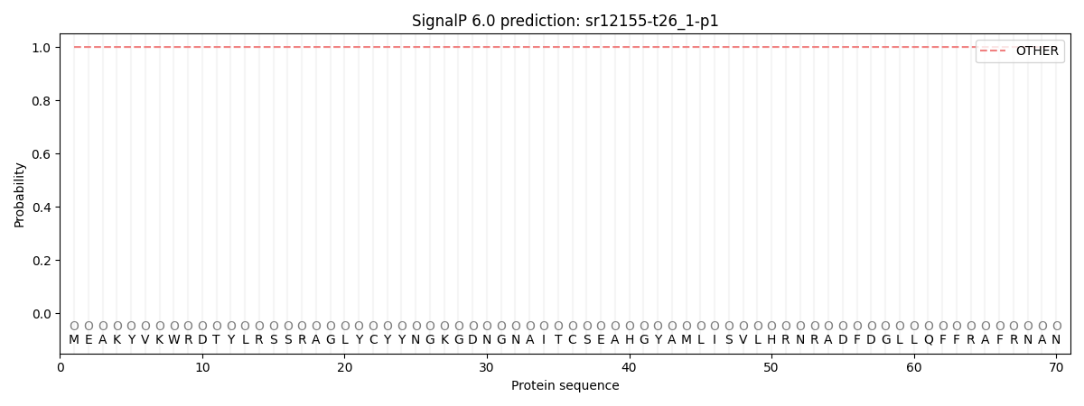
This protein is predicted as OTHER

| Other | SP_Sec_SPI | CS Position |
|---|---|---|
| 1.000067 | 0.000000 |
