You are browsing environment: FUNGIDB
CAZyme Information: jhhlp_001580-t41_1-p1
You are here: Home > Sequence: jhhlp_001580-t41_1-p1
Basic Information |
Genomic context |
Full Sequence |
Enzyme annotations |
CAZy signature domains |
CDD domains |
CAZyme hits |
PDB hits |
Swiss-Prot hits |
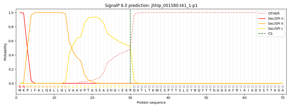
SignalP and Lipop annotations |
TMHMM annotations
Basic Information help
| Species | Lomentospora prolificans | |||||||||||
|---|---|---|---|---|---|---|---|---|---|---|---|---|
| Lineage | Ascomycota; Sordariomycetes; ; Microascaceae; Lomentospora; Lomentospora prolificans | |||||||||||
| CAZyme ID | jhhlp_001580-t41_1-p1 | |||||||||||
| CAZy Family | AA7 | |||||||||||
| CAZyme Description | hypothetical protein | |||||||||||
| CAZyme Property |
|
|||||||||||
| Genome Property |
|
|||||||||||
| Gene Location | ||||||||||||
CAZyme Signature Domains help
| Family | Start | End | Evalue | family coverage |
|---|---|---|---|---|
| AA8 | 39 | 211 | 2.8e-47 | 0.9505494505494505 |
CDD Domains download full data without filtering help
| Cdd ID | Domain | E-Value | qStart | qEnd | sStart | sEnd | Domain Description |
|---|---|---|---|---|---|---|---|
| 406418 | CDH-cyt | 1.79e-55 | 37 | 212 | 1 | 176 | Cytochrome domain of cellobiose dehydrogenase. CDH-cyt is the cytochrome domain, at the N-terminus, of cellobiose dehydrogenase. CDH-cyt folds as a beta sandwich with the topology of the antibody Fab V(H) domain and binds iron. The haem iron is ligated by Met83 and His181 in UniProtKB:Q01738. |
| 187688 | CDH_like_cytochrome | 8.67e-45 | 37 | 204 | 2 | 166 | Heme-binding cytochrome domain of fungal cellobiose dehydrogenases. Cellobiose dehydrogenase (CellobioseDH or CDH) is an extracellular fungal oxidoreductase that degrades both lignin and cellulose. Specifically, CDHs oxidize cellobiose, cellodextrins, and lactose to corresponding lactones, utilizing a variety of electron acceptors. Class-II CDHs are monomeric hemoflavoenzymes that are comprised of a b-type cytochrome domain linked to a large flavodehydrogenase domain. The cytochrome domain of CDH and related enzymes, which this model describes, folds as a beta sandwich and complexes a heme molecule. It is found at the N-terminus of this family of enzymes, and belongs to the DOMON domain superfamily, a ligand-interacting motif found in all three kingdoms of life. |
| 187689 | DOMON_DOH | 2.31e-06 | 72 | 197 | 20 | 134 | DOMON-like domain of copper-dependent monooxygenases and related proteins. This diverse family characterizes DOMON domains found in dopamine beta-hydroxylase (DBH), monooxygenase X (MOX), and various other proteins, some of which contain DOMON domains exclusively; the family is not restricted to eukaryotes. DBH is a membrane-bound enzyme that converts dopamine to L-norepinephrine, and plays a central role in the metabolism of catecholamine neurotransmitters. DOMON domains were initially thought to confer protein-protein interactions. They were subsequently found as a heme-binding motif in cellobiose dehydrogenase, an extracellular fungal oxidoreductase that degrades both lignin and cellulose, and in ethylbenzene dehydrogenase, an enzyme that aids in the anaerobic degradation of hydrocarbons. The domain interacts with sugars in the type 9 carbohydrate binding modules (CBM9), which are present in a variety of glycosyl hydrolases, and it can also be found at the N-terminus of sensor histidine kinases. |
| 397434 | DOMON | 7.18e-04 | 72 | 175 | 15 | 118 | DOMON domain. The DOMON (named after dopamine beta-monooxygenase N-terminal) domain is 110-125 residues long. It is predicted to form an all beta fold with up to 11 strands and is secreted to the extracellular compartment. The beta-strand folding produces a hydrophobic pocket which appears to bind soluble haem. This is consistent with the predominant architectures where the protein is associated with cytochromes or enzymatic domains whose activity involves redox or electron transfer reactions potentially as a direct participant in the electron transfer process. The DOMON domain superfamily, of which this is just one member, shows (1) multiple hydrophobic residues that contribute to the hydrophobic core of the strands of the beta-sandwich, and small residues found at the boundaries of strands and loops, (2) a strongly conserved charged residue (usually arginine/lysine) at the end of strand 9, which possibly stabilizes the loop between 9 and 10, and (3) a polar residue (usually histidine, lysine or arginine), that interacts or coordinates with ligands. The suggested superfamily includes both haem- and sugar-binding members: the haem-binding families being the ethyl-Benzoate dehydrogenase family EB_dh, pfam09459, the cellobiose dehydrogenase family CBDH and this family, and the sugar-binding families being the xylanases, CBM_4_9, pfam02018. The common feature of the superfamily is the 11-beta-strand structure, although the first and eleventh strands are not well conserved either within families or between families. |
CAZyme Hits help
| Hit ID | E-Value | Query Start | Query End | Hit Start | Hit End |
|---|---|---|---|---|---|
| 4.84e-47 | 16 | 220 | 24 | 227 | |
| 4.58e-46 | 20 | 216 | 27 | 217 | |
| 1.27e-37 | 24 | 221 | 22 | 211 | |
| 5.05e-37 | 24 | 221 | 22 | 211 | |
| 5.53e-36 | 17 | 218 | 27 | 219 |
PDB Hits download full data without filtering help
| Hit ID | E-Value | Query Start | Query End | Hit Start | Hit End | Description |
|---|---|---|---|---|---|---|
| 1.73e-20 | 76 | 206 | 47 | 173 | Chain A, CELLOBIOSE DEHYDROGENASE [Phanerodontia chrysosporium],1D7B_B Chain B, CELLOBIOSE DEHYDROGENASE [Phanerodontia chrysosporium],1D7D_A Chain A, CELLOBIOSE DEHYDROGENASE [Phanerodontia chrysosporium],1D7D_B Chain B, CELLOBIOSE DEHYDROGENASE [Phanerodontia chrysosporium] |
|
| 1.89e-20 | 76 | 206 | 47 | 173 | Chain A, CELLOBIOSE DEHYDROGENASE [Phanerodontia chrysosporium],1D7C_B Chain B, CELLOBIOSE DEHYDROGENASE [Phanerodontia chrysosporium] |
|
| 1.80e-19 | 76 | 206 | 47 | 173 | Chain A, Cellobiose dehydrogenase [Phanerodontia chrysosporium],1PL3_B Chain B, Cellobiose dehydrogenase [Phanerodontia chrysosporium] |
|
| 2.36e-14 | 31 | 202 | 1 | 170 | Chain A, Extracellular PQQ-dependent sugar dehydrogenase [Coprinopsis cinerea] |
|
| 1.38e-12 | 51 | 189 | 29 | 165 | Chain A, Cellobiose dehydrogenase [Thermothelomyces myriococcoides],4QI3_B Chain B, Cellobiose dehydrogenase [Thermothelomyces myriococcoides] |
Swiss-Prot Hits download full data without filtering help
| Hit ID | E-Value | Query Start | Query End | Hit Start | Hit End | Description |
|---|---|---|---|---|---|---|
| 3.14e-18 | 76 | 206 | 65 | 191 | Cellobiose dehydrogenase OS=Phanerodontia chrysosporium OX=2822231 GN=CDH-1 PE=1 SV=1 |
SignalP and Lipop Annotations help
This protein is predicted as SP

| Other | SP_Sec_SPI | CS Position |
|---|---|---|
| 0.000221 | 0.999750 | CS pos: 30-31. Pr: 0.5168 |
