You are browsing environment: FUNGIDB
CAZyme Information: TRIREDRAFT_121491-t26_1-p1
You are here: Home > Sequence: TRIREDRAFT_121491-t26_1-p1
Basic Information |
Genomic context |
Full Sequence |
Enzyme annotations |
CAZy signature domains |
CDD domains |
CAZyme hits |
PDB hits |
Swiss-Prot hits |
SignalP and Lipop annotations |
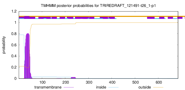
TMHMM annotations
Basic Information help
| Species | Trichoderma reesei | |||||||||||
|---|---|---|---|---|---|---|---|---|---|---|---|---|
| Lineage | Ascomycota; Sordariomycetes; ; Hypocreaceae; Trichoderma; Trichoderma reesei | |||||||||||
| CAZyme ID | TRIREDRAFT_121491-t26_1-p1 | |||||||||||
| CAZy Family | CBM21 | |||||||||||
| CAZyme Description | glycosyltransferase family 4 | |||||||||||
| CAZyme Property |
|
|||||||||||
| Genome Property |
|
|||||||||||
| Gene Location | ||||||||||||
Enzyme Prediction help
| EC | 2.4.1.231:3 |
|---|
CDD Domains download full data without filtering help
| Cdd ID | Domain | E-Value | qStart | qEnd | sStart | sEnd | Domain Description |
|---|---|---|---|---|---|---|---|
| 340823 | GT4_trehalose_phosphorylase | 0.0 | 224 | 643 | 1 | 378 | trehalose phosphorylase and similar proteins. Trehalose phosphorylase (TP) reversibly catalyzes trehalose synthesis and degradation from alpha-glucose-1-phosphate (alpha-Glc-1-P) and glucose. The catalyzing activity includes the phosphorolysis of trehalose, which produce alpha-Glc-1-P and glucose, and the subsequent synthesis of trehalose. This family is most closely related to the GT4 family of glycosyltransferases. |
| 395425 | Glycos_transf_1 | 1.61e-21 | 449 | 620 | 2 | 156 | Glycosyl transferases group 1. Mutations in this domain of PIGA lead to disease (Paroxysmal Nocturnal haemoglobinuria). Members of this family transfer activated sugars to a variety of substrates, including glycogen, Fructose-6-phosphate and lipopolysaccharides. Members of this family transfer UDP, ADP, GDP or CMP linked sugars. The eukaryotic glycogen synthases may be distant members of this family. |
| 223515 | RfaB | 2.26e-21 | 224 | 619 | 4 | 353 | Glycosyltransferase involved in cell wall bisynthesis [Cell wall/membrane/envelope biogenesis]. |
| 340831 | GT4_PimA-like | 1.06e-20 | 447 | 620 | 190 | 345 | phosphatidyl-myo-inositol mannosyltransferase. This family is most closely related to the GT4 family of glycosyltransferases and named after PimA in Propionibacterium freudenreichii, which is involved in the biosynthesis of phosphatidyl-myo-inositol mannosides (PIM) which are early precursors in the biosynthesis of lipomannans (LM) and lipoarabinomannans (LAM), and catalyzes the addition of a mannosyl residue from GDP-D-mannose (GDP-Man) to the position 2 of the carrier lipid phosphatidyl-myo-inositol (PI) to generate a phosphatidyl-myo-inositol bearing an alpha-1,2-linked mannose residue (PIM1). Glycosyltransferases catalyze the transfer of sugar moieties from activated donor molecules to specific acceptor molecules, forming glycosidic bonds. The acceptor molecule can be a lipid, a protein, a heterocyclic compound, or another carbohydrate residue. This group of glycosyltransferases is most closely related to the previously defined glycosyltransferase family 1 (GT1). The members of this family may transfer UDP, ADP, GDP, or CMP linked sugars. The diverse enzymatic activities among members of this family reflect a wide range of biological functions. The protein structure available for this family has the GTB topology, one of the two protein topologies observed for nucleotide-sugar-dependent glycosyltransferases. GTB proteins have distinct N- and C- terminal domains each containing a typical Rossmann fold. The two domains have high structural homology despite minimal sequence homology. The large cleft that separates the two domains includes the catalytic center and permits a high degree of flexibility. The members of this family are found mainly in certain bacteria and archaea. |
| 340830 | GT4_sucrose_synthase | 1.31e-20 | 440 | 619 | 210 | 379 | sucrose-phosphate synthase and similar proteins. This family is most closely related to the GT4 family of glycosyltransferases. The sucrose-phosphate synthases in this family may be unique to plants and photosynthetic bacteria. This enzyme catalyzes the synthesis of sucrose 6-phosphate from fructose 6-phosphate and uridine 5'-diphosphate-glucose, a key regulatory step of sucrose metabolism. The activity of this enzyme is regulated by phosphorylation and moderated by the concentration of various metabolites and light. |
CAZyme Hits help
| Hit ID | E-Value | Query Start | Query End | Hit Start | Hit End |
|---|---|---|---|---|---|
| 0.0 | 1 | 685 | 1 | 680 | |
| 0.0 | 7 | 682 | 7 | 684 | |
| 0.0 | 1 | 682 | 1 | 679 | |
| 0.0 | 119 | 682 | 2 | 565 | |
| 0.0 | 159 | 685 | 1 | 527 |
PDB Hits download full data without filtering help
| Hit ID | E-Value | Query Start | Query End | Hit Start | Hit End | Description |
|---|---|---|---|---|---|---|
| 2.73e-39 | 196 | 623 | 16 | 394 | Crystal structure of trehalose synthase TreT from P.horikoshi [Pyrococcus horikoshii],2X6Q_B Crystal structure of trehalose synthase TreT from P.horikoshi [Pyrococcus horikoshii],2X6R_A Crystal structure of trehalose synthase TreT from P.horikoshi produced by soaking in trehalose [Pyrococcus horikoshii],2X6R_B Crystal structure of trehalose synthase TreT from P.horikoshi produced by soaking in trehalose [Pyrococcus horikoshii] |
|
| 1.76e-38 | 196 | 623 | 16 | 394 | Crystal structure of trehalose synthase TreT mutant E326A from P. horikoshii in complex with UDPG [Pyrococcus horikoshii],2XA2_B Crystal structure of trehalose synthase TreT mutant E326A from P. horikoshii in complex with UDPG [Pyrococcus horikoshii],2XA9_A Crystal structure of trehalose synthase TreT mutant E326A from P. horikoshii in complex with UDPG [Pyrococcus horikoshii],2XA9_B Crystal structure of trehalose synthase TreT mutant E326A from P. horikoshii in complex with UDPG [Pyrococcus horikoshii],2XMP_A Crystal structure of trehalose synthase TreT mutant E326A from P. horishiki in complex with UDP [Pyrococcus horikoshii],2XMP_B Crystal structure of trehalose synthase TreT mutant E326A from P. horishiki in complex with UDP [Pyrococcus horikoshii] |
|
| 3.90e-37 | 196 | 591 | 16 | 364 | Crystal structure of trehalose synthase TreT from P.horikoshii (Seleno derivative) [Pyrococcus horikoshii],2XA1_B Crystal structure of trehalose synthase TreT from P.horikoshii (Seleno derivative) [Pyrococcus horikoshii] |
|
| 1.53e-31 | 209 | 623 | 15 | 375 | Chain AAA, Trehalose phosphorylase/synthase [Thermoproteus uzoniensis],6ZJ7_AAA Chain AAA, Trehalose phosphorylase/synthase [Thermoproteus uzoniensis 768-20],6ZJH_AAA Chain AAA, Trehalose phosphorylase/synthase [Thermoproteus uzoniensis 768-20],6ZMZ_AAA Chain AAA, Trehalose phosphorylase/synthase [Thermoproteus uzoniensis],6ZN1_AAA Chain AAA, Trehalose phosphorylase/synthase [Thermoproteus uzoniensis] |
|
| 1.22e-07 | 447 | 638 | 198 | 371 | Chain A, Glycosyltransferase [Staphylococcus aureus subsp. aureus CN1] |
Swiss-Prot Hits download full data without filtering help
| Hit ID | E-Value | Query Start | Query End | Hit Start | Hit End | Description |
|---|---|---|---|---|---|---|
| 4.22e-212 | 34 | 680 | 35 | 725 | Trehalose phosphorylase OS=Grifola frondosa OX=5627 PE=1 SV=1 |
|
| 9.73e-210 | 34 | 681 | 39 | 733 | Trehalose phosphorylase OS=Pleurotus pulmonarius OX=28995 GN=TP PE=2 SV=1 |
|
| 2.19e-203 | 34 | 681 | 39 | 745 | Trehalose phosphorylase OS=Pleurotus sajor-caju OX=50053 GN=TP PE=1 SV=1 |
|
| 7.39e-39 | 196 | 623 | 15 | 393 | Trehalose synthase OS=Pyrococcus horikoshii (strain ATCC 700860 / DSM 12428 / JCM 9974 / NBRC 100139 / OT-3) OX=70601 GN=treT PE=1 SV=2 |
|
| 9.52e-39 | 196 | 591 | 14 | 362 | Trehalose synthase OS=Thermococcus litoralis (strain ATCC 51850 / DSM 5473 / JCM 8560 / NS-C) OX=523849 GN=treT PE=1 SV=1 |
SignalP and Lipop Annotations help
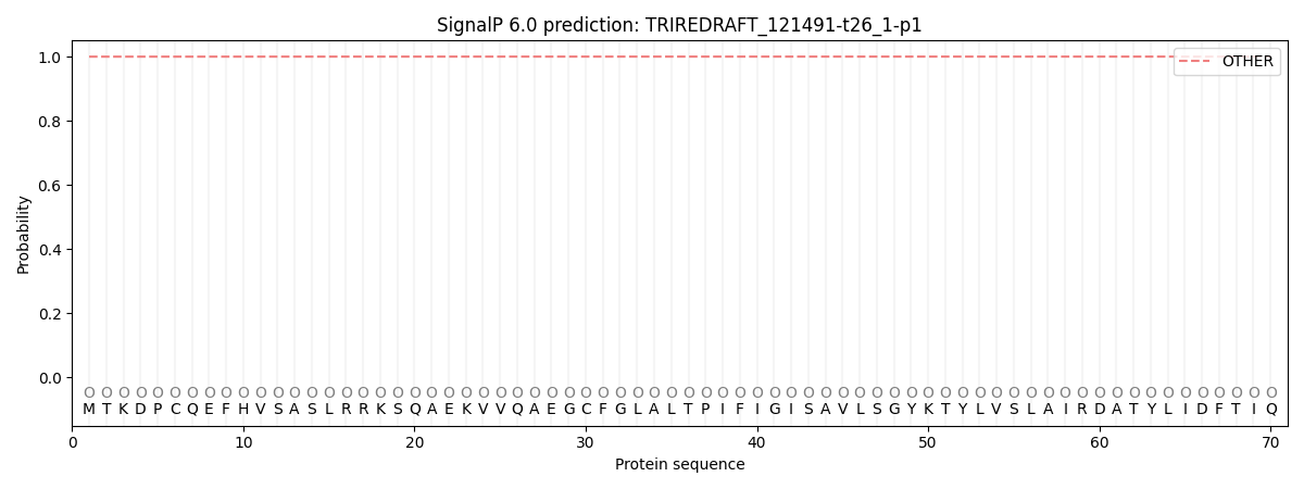
This protein is predicted as OTHER

| Other | SP_Sec_SPI | CS Position |
|---|---|---|
| 0.999883 | 0.000115 |

