You are browsing environment: FUNGIDB
CAZyme Information: TPX50376.1
You are here: Home > Sequence: TPX50376.1
Basic Information |
Genomic context |
Full Sequence |
Enzyme annotations |
CAZy signature domains |
CDD domains |
CAZyme hits |
PDB hits |
Swiss-Prot hits |
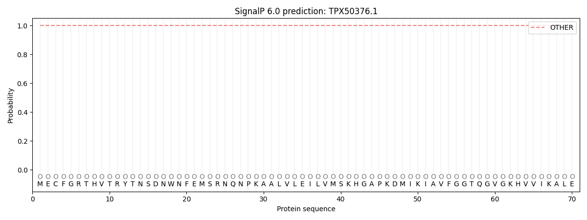
SignalP and Lipop annotations |
TMHMM annotations
Basic Information help
| Species | Synchytrium endobioticum | |||||||||||
|---|---|---|---|---|---|---|---|---|---|---|---|---|
| Lineage | Chytridiomycota; Chytridiomycetes; ; Synchytriaceae; Synchytrium; Synchytrium endobioticum | |||||||||||
| CAZyme ID | TPX50376.1 | |||||||||||
| CAZy Family | GT2 | |||||||||||
| CAZyme Description | glucan endo-1,3-beta-D-glucosidase | |||||||||||
| CAZyme Property |
|
|||||||||||
| Genome Property |
|
|||||||||||
| Gene Location | Start: 41845; End:44198 Strand: + | |||||||||||
Full Sequence Download help
| MECFGRTHVT RYTNSDNWNF EMSRNQNPKA ALVLEILVMS KHGAPKDMIK IAVFGGTQGV | 60 |
| GKHVVIKALE CHHFVTVLAR SPKKLADVVV NRHKLKIIQG DILHDPAAVD QVVDGQDVVI | 120 |
| VSLGTTGVKK GPQAKVCSFG QKVINDAMKA RGVKRLIVVT SIGVGDSIKH VSYSAYFFIK | 180 |
| FVIYKAIADK EVQEDLVKSS GLDWTILRPT GLLDQPPRSL RYDYGEDLNG SSIARAHVAE | 240 |
| ICLEVIPKPH YGRIYALTLL HTLHFRHGQD MYAHCISLIL LLICCSQVKP DLYGINYSPR | 300 |
| RSLFACPTHA MITSDLQLLR NMTTRIKTFS LIDSGSGQAS ATCNFGETIL KAAVPLGFRV | 360 |
| TLGMEFRGAD IDAMFQREVR ELERLSYAYP NLFTVVEAIV VGSETLYRGE ETQQTLSQRV | 420 |
| TTVRTILHSK NLRVPITAAD IPPPFYGDIL IAAVDFIMIN VYPYWEGHAV ASAPYSQMAA | 480 |
| VHALRERTSK NVVLGECGWP TAGSTNTDAE ASIQNLEWYY KQWVCTARKN GVEYFLFEAF | 540 |
| DEDWKSDEHA GVEKHWGLFD LNRQPKSTVF LNPVDCSP | 578 |
CAZyme Signature Domains help
| Family | Start | End | Evalue | family coverage |
|---|---|---|---|---|
| GH17 | 385 | 566 | 7.4e-34 | 0.7331189710610932 |
CDD Domains download full data without filtering help
| Cdd ID | Domain | E-Value | qStart | qEnd | sStart | sEnd | Domain Description |
|---|---|---|---|---|---|---|---|
| 227625 | Scw11 | 8.57e-53 | 294 | 563 | 47 | 305 | Exo-beta-1,3-glucanase, GH17 family [Carbohydrate transport and metabolism]. |
| 404360 | NAD_binding_10 | 4.73e-47 | 55 | 249 | 1 | 183 | NAD(P)H-binding. |
| 187555 | BVR-B_like_SDR_a | 7.83e-43 | 50 | 239 | 1 | 188 | biliverdin IX beta reductase (BVR-B, aka flavin reductase)-like proteins; atypical (a) SDRs. Human BVR-B catalyzes pyridine nucleotide-dependent production of bilirubin-IX beta during fetal development; in the adult BVR-B has flavin and ferric reductase activities. Human BVR-B catalyzes the reduction of FMN, FAD, and riboflavin. Recognition of flavin occurs mostly by hydrophobic interactions, accounting for the broad substrate specificity. Atypical SDRs are distinct from classical SDRs. BVR-B does not share the key catalytic triad, or conserved tyrosine typical of SDRs. The glycine-rich NADP-binding motif of BVR-B is GXXGXXG, which is similar but not identical to the pattern seen in extended SDRs. Atypical SDRs generally lack the catalytic residues characteristic of the SDRs, and their glycine-rich NAD(P)-binding motif is often different from the forms normally seen in classical or extended SDRs. Atypical SDRs include biliverdin IX beta reductase (BVR-B,aka flavin reductase), NMRa (a negative transcriptional regulator of various fungi), progesterone 5-beta-reductase like proteins, phenylcoumaran benzylic ether and pinoresinol-lariciresinol reductases, phenylpropene synthases, eugenol synthase, triphenylmethane reductase, isoflavone reductases, and others. SDRs are a functionally diverse family of oxidoreductases that have a single domain with a structurally conserved Rossmann fold, an NAD(P)(H)-binding region, and a structurally diverse C-terminal region. Sequence identity between different SDR enzymes is typically in the 15-30% range; they catalyze a wide range of activities including the metabolism of steroids, cofactors, carbohydrates, lipids, aromatic compounds, and amino acids, and act in redox sensing. Classical SDRs have an TGXXX[AG]XG cofactor binding motif and a YXXXK active site motif, with the Tyr residue of the active site motif serving as a critical catalytic residue (Tyr-151, human 15-hydroxyprostaglandin dehydrogenase numbering). In addition to the Tyr and Lys, there is often an upstream Ser and/or an Asn, contributing to the active site; while substrate binding is in the C-terminal region, which determines specificity. The standard reaction mechanism is a 4-pro-S hydride transfer and proton relay involving the conserved Tyr and Lys, a water molecule stabilized by Asn, and nicotinamide. In addition to the Rossmann fold core region typical of all SDRs, extended SDRs have a less conserved C-terminal extension of approximately 100 amino acids, and typically have a TGXXGXXG cofactor binding motif. Complex (multidomain) SDRs such as ketoreductase domains of fatty acid synthase have a GGXGXXG NAD(P)-binding motif and an altered active site motif (YXXXN). Fungal type ketoacyl reductases have a TGXXXGX(1-2)G NAD(P)-binding motif. |
| 187554 | SDR_a5 | 4.44e-32 | 50 | 244 | 1 | 185 | atypical (a) SDRs, subgroup 5. This subgroup contains atypical SDRs, some of which are identified as putative NAD(P)-dependent epimerases, one as a putative NAD-dependent epimerase/dehydratase. Atypical SDRs are distinct from classical SDRs. Members of this subgroup have a glycine-rich NAD(P)-binding motif that is very similar to the extended SDRs, GXXGXXG, and binds NADP. Generally, this subgroup has poor conservation of the active site tetrad; however, individual sequences do contain matches to the YXXXK active site motif, the upstream Ser, and there is a highly conserved Asp in place of the usual active site Asn throughout the subgroup. Atypical SDRs generally lack the catalytic residues characteristic of the SDRs, and their glycine-rich NAD(P)-binding motif is often different from the forms normally seen in classical or extended SDRs. Atypical SDRs include biliverdin IX beta reductase (BVR-B,aka flavin reductase), NMRa (a negative transcriptional regulator of various fungi), progesterone 5-beta-reductase like proteins, phenylcoumaran benzylic ether and pinoresinol-lariciresinol reductases, phenylpropene synthases, eugenol synthase, triphenylmethane reductase, isoflavone reductases, and others. SDRs are a functionally diverse family of oxidoreductases that have a single domain with a structurally conserved Rossmann fold, an NAD(P)(H)-binding region, and a structurally diverse C-terminal region. Sequence identity between different SDR enzymes is typically in the 15-30% range; they catalyze a wide range of activities including the metabolism of steroids, cofactors, carbohydrates, lipids, aromatic compounds, and amino acids, and act in redox sensing. Classical SDRs have an TGXXX[AG]XG cofactor binding motif and a YXXXK active site motif, with the Tyr residue of the active site motif serving as a critical catalytic residue (Tyr-151, human 15-hydroxyprostaglandin dehydrogenase numbering). In addition to the Tyr and Lys, there is often an upstream Ser and/or an Asn, contributing to the active site; while substrate binding is in the C-terminal region, which determines specificity. The standard reaction mechanism is a 4-pro-S hydride transfer and proton relay involving the conserved Tyr and Lys, a water molecule stabilized by Asn, and nicotinamide. In addition to the Rossmann fold core region typical of all SDRs, extended SDRs have a less conserved C-terminal extension of approximately 100 amino acids, and typically have a TGXXGXXG cofactor binding motif. Complex (multidomain) SDRs such as ketoreductase domains of fatty acid synthase have a GGXGXXG NAD(P)-binding motif and an altered active site motif (YXXXN). Fungal type ketoacyl reductases have a TGXXXGX(1-2)G NAD(P)-binding motif. |
| 366033 | Glyco_hydro_17 | 9.21e-20 | 450 | 566 | 171 | 304 | Glycosyl hydrolases family 17. |
CAZyme Hits help
| Hit ID | E-Value | Query Start | Query End | Hit Start | Hit End |
|---|---|---|---|---|---|
| QLQ77781.1|GH17 | 2.39e-29 | 281 | 572 | 10 | 307 |
| CCD23099.1|GH17 | 2.60e-29 | 281 | 569 | 13 | 307 |
| ABP48761.2|GH17 | 8.87e-29 | 306 | 559 | 39 | 297 |
| CCE61637.1|GH17 | 8.87e-29 | 306 | 559 | 39 | 297 |
| QLL30211.1|GH17 | 1.56e-28 | 281 | 569 | 10 | 304 |
PDB Hits download full data without filtering help
| Hit ID | E-Value | Query Start | Query End | Hit Start | Hit End | Description |
|---|---|---|---|---|---|---|
| 4WTP_A | 4.23e-33 | 293 | 569 | 38 | 294 | Crystal structure of glycoside hydrolase family 17 beta-1,3-glucanosyltransferase from Rhizomucor miehei [Rhizomucor miehei CAU432] |
| 4WTR_A | 2.81e-32 | 293 | 569 | 38 | 294 | Active-site mutant of Rhizomucor miehei beta-1,3-glucanosyltransferase in complex with laminaribiose [Rhizomucor miehei CAU432],4WTS_A Active-site mutant of Rhizomucor miehei beta-1,3-glucanosyltransferase in complex with laminaritriose [Rhizomucor miehei CAU432] |
| 3QVO_A | 3.19e-10 | 51 | 249 | 26 | 211 | Structure of a Rossmann-fold NAD(P)-binding family protein from Shigella flexneri. [Shigella flexneri 2a str. 2457T] |
| 4JGB_A | 7.61e-10 | 45 | 225 | 2 | 172 | Crystal Structure of Putative exported protein from Burkholderia pseudomallei [Burkholderia pseudomallei K96243],4JGB_B Crystal Structure of Putative exported protein from Burkholderia pseudomallei [Burkholderia pseudomallei K96243] |
| 5FFQ_A | 9.39e-07 | 143 | 214 | 89 | 155 | ChuY: An Anaerobillin Reductase from Escherichia coli O157:H7 [Escherichia coli O157:H7],5FFQ_B ChuY: An Anaerobillin Reductase from Escherichia coli O157:H7 [Escherichia coli O157:H7] |
Swiss-Prot Hits download full data without filtering help
| Hit ID | E-Value | Query Start | Query End | Hit Start | Hit End | Description |
|---|---|---|---|---|---|---|
| sp|D4B2W4|BGL2_ARTBC | 9.59e-32 | 272 | 566 | 3 | 296 | Glucan 1,3-beta-glucosidase ARB_02797 OS=Arthroderma benhamiae (strain ATCC MYA-4681 / CBS 112371) OX=663331 GN=ARB_02797 PE=1 SV=1 |
| sp|O13990|BGL2_SCHPO | 5.76e-28 | 314 | 566 | 63 | 313 | Glucan 1,3-beta-glucosidase OS=Schizosaccharomyces pombe (strain 972 / ATCC 24843) OX=284812 GN=bgl2 PE=2 SV=4 |
| sp|P15703|BGL2_YEAST | 1.25e-27 | 306 | 559 | 40 | 298 | Glucan 1,3-beta-glucosidase OS=Saccharomyces cerevisiae (strain ATCC 204508 / S288c) OX=559292 GN=BGL2 PE=1 SV=1 |
| sp|P43070|BGL2_CANAX | 1.15e-23 | 306 | 569 | 35 | 303 | Glucan 1,3-beta-glucosidase OS=Candida albicans OX=5476 GN=BGL2 PE=3 SV=1 |
| sp|Q5AMT2|BGL2_CANAL | 1.56e-23 | 306 | 569 | 35 | 303 | Glucan 1,3-beta-glucosidase BGL2 OS=Candida albicans (strain SC5314 / ATCC MYA-2876) OX=237561 GN=BGL2 PE=1 SV=2 |
SignalP and Lipop Annotations help
This protein is predicted as OTHER

| Other | SP_Sec_SPI | CS Position |
|---|---|---|
| 1.000079 | 0.000000 |
