You are browsing environment: FUNGIDB
CAZyme Information: SPRG_19057-t26_1-p1
You are here: Home > Sequence: SPRG_19057-t26_1-p1
Basic Information |
Genomic context |
Full Sequence |
Enzyme annotations |
CAZy signature domains |
CDD domains |
CAZyme hits |
PDB hits |
Swiss-Prot hits |
SignalP and Lipop annotations |
TMHMM annotations
Basic Information help
| Species | Saprolegnia parasitica | |||||||||||
|---|---|---|---|---|---|---|---|---|---|---|---|---|
| Lineage | Oomycota; NA; ; Saprolegniaceae; Saprolegnia; Saprolegnia parasitica | |||||||||||
| CAZyme ID | SPRG_19057-t26_1-p1 | |||||||||||
| CAZy Family | GT41 | |||||||||||
| CAZyme Description | hypothetical protein | |||||||||||
| CAZyme Property |
|
|||||||||||
| Genome Property |
|
|||||||||||
| Gene Location | Start: 702569; End:705865 Strand: + | |||||||||||
Full Sequence Download help
| MYRASAKVSS AHNDGIWSTV WTSRNQILSG SVDEVVKSWD ASSTEDNAIL PVVKQFPGHV | 60 |
| LGTLGVAATK DGRRAATSSL DCQVRILNLE TGGVEKTIDT GAGETWQVAY SPDGAFVATG | 120 |
| SQQGKINIIN VHDEKVVQSI VVEEEKPAKA SSSSDAKGKF VLSVAYSPDG KHLACSTFDG | 180 |
| LVAIYDIESG KQLQKYQDRT KPVRSIAYSP DGSFLLAASD DMHVNIYDVA HSSMVASVAG | 240 |
| HISWVLSVAC SPDGKHFATG GGDRKVKIWD LAAKSCLYTF ECHTDQVWSV AYNETGSRLV | 300 |
| SGGDDALLQL YEVSTASLAH GHIQADIRAG RVPSRGVNLG GWLVAEHWMT ASSSIWNGVP | 360 |
| ADRASQGEYH AMSVLGHNVG DAAFESHRRS FLTQADIAQI GAAGLNTVRV PIGYWIRGCG | 420 |
| MLTGPLFQQC SVFAPGGLKY LDMLIQQWAR AANVAVLISI HGAPGSQNGN DHSAAVMKGR | 480 |
| VDWPNDPVNV KVTRDLVLFL VQRYKNEQAF LGIGLLNEPD GHMNSHVLFS YYTASYNDVR | 540 |
| SISDCILTMM PRLYNQYAGN GDAMGDFGKG MQNVWIEWHP YLIWGYESYS EAMLLNQGID | 600 |
| SIAKNIANWR GHPLYFGEWS VVTPSNTFSD PNALASFRRK LVTVMNTAQG WAYWTWRADG | 660 |
| DGYGHKWSLR DLLRRMQYPI VRTAANAVGS SPPLSILQSN GVGLSLLTQT QSLAADPAWI | 720 |
| AAMGPLVAER WSYNPSTQQL QSLTSGNCLD AYFDNGLQRH VIHSYSCDGT NANQKWTLVN | 780 |
| HQLSNKGLCL ALVDAAINTD LVANRRLVTC DSASTAQFFT LGIAVARLRI ASLASLVLSA | 840 |
| SLTFQAPSAR DTSQLWLFNH LDYTVMNQGS GQCLDAYEAK VGGAVHLYAC SPGNVNQLWR | 900 |
| YNPLTRQLQH MGHAGYCLDV YGPPHLNTCY AMGNAAMWAQ ALQLEWISYP ALTGGYAAVT | 960 |
| SS | 962 |
CAZyme Signature Domains help
| Family | Start | End | Evalue | family coverage |
|---|---|---|---|---|
| GH5 | 385 | 661 | 6.7e-39 | 0.9711191335740073 |
CDD Domains download full data without filtering help
| Cdd ID | Domain | E-Value | qStart | qEnd | sStart | sEnd | Domain Description |
|---|---|---|---|---|---|---|---|
| 238121 | WD40 | 3.10e-54 | 12 | 312 | 8 | 289 | WD40 domain, found in a number of eukaryotic proteins that cover a wide variety of functions including adaptor/regulatory modules in signal transduction, pre-mRNA processing and cytoskeleton assembly; typically contains a GH dipeptide 11-24 residues from its N-terminus and the WD dipeptide at its C-terminus and is 40 residues long, hence the name WD40; between GH and WD lies a conserved core; serves as a stable propeller-like platform to which proteins can bind either stably or reversibly; forms a propeller-like structure with several blades where each blade is composed of a four-stranded anti-parallel b-sheet; instances with few detectable copies are hypothesized to form larger structures by dimerization; each WD40 sequence repeat forms the first three strands of one blade and the last strand in the next blade; the last C-terminal WD40 repeat completes the blade structure of the first WD40 repeat to create the closed ring propeller-structure; residues on the top and bottom surface of the propeller are proposed to coordinate interactions with other proteins and/or small ligands; 7 copies of the repeat are present in this alignment. |
| 238121 | WD40 | 2.03e-48 | 53 | 322 | 2 | 260 | WD40 domain, found in a number of eukaryotic proteins that cover a wide variety of functions including adaptor/regulatory modules in signal transduction, pre-mRNA processing and cytoskeleton assembly; typically contains a GH dipeptide 11-24 residues from its N-terminus and the WD dipeptide at its C-terminus and is 40 residues long, hence the name WD40; between GH and WD lies a conserved core; serves as a stable propeller-like platform to which proteins can bind either stably or reversibly; forms a propeller-like structure with several blades where each blade is composed of a four-stranded anti-parallel b-sheet; instances with few detectable copies are hypothesized to form larger structures by dimerization; each WD40 sequence repeat forms the first three strands of one blade and the last strand in the next blade; the last C-terminal WD40 repeat completes the blade structure of the first WD40 repeat to create the closed ring propeller-structure; residues on the top and bottom surface of the propeller are proposed to coordinate interactions with other proteins and/or small ligands; 7 copies of the repeat are present in this alignment. |
| 225201 | WD40 | 4.21e-39 | 7 | 315 | 149 | 443 | WD40 repeat [General function prediction only]. |
| 293791 | 7WD40 | 7.62e-35 | 26 | 311 | 12 | 293 | WD40 repeats in seven bladed beta propellers. The WD40 repeat is found in a number of eukaryotic proteins that cover a wide variety of functions including adaptor/regulatory modules in signal transduction, pre-mRNA processing, and cytoskeleton assembly. It typically contains a GH dipeptide 11-24 residues from its N-terminus and the WD dipeptide at its C-terminus and is 40 residues long, hence the name WD40. Between the GH and WD dipeptides lies a conserved core. It forms a propeller-like structure with several blades where each blade is composed of a four-stranded anti-parallel beta-sheet. The WD40 sequence repeat originally described in literature forms the first three strands of one blade and the last strand in the next blade. The C-terminal WD40 repeat completes the blade structure of the N-terminal WD40 repeat to create the closed ring propeller-structure. The residues on the top and bottom surface of the propeller are proposed to coordinate interactions with other proteins and/or small ligands, allowing them to bind either stably or reversibly. |
| 293791 | 7WD40 | 1.47e-33 | 106 | 315 | 2 | 204 | WD40 repeats in seven bladed beta propellers. The WD40 repeat is found in a number of eukaryotic proteins that cover a wide variety of functions including adaptor/regulatory modules in signal transduction, pre-mRNA processing, and cytoskeleton assembly. It typically contains a GH dipeptide 11-24 residues from its N-terminus and the WD dipeptide at its C-terminus and is 40 residues long, hence the name WD40. Between the GH and WD dipeptides lies a conserved core. It forms a propeller-like structure with several blades where each blade is composed of a four-stranded anti-parallel beta-sheet. The WD40 sequence repeat originally described in literature forms the first three strands of one blade and the last strand in the next blade. The C-terminal WD40 repeat completes the blade structure of the N-terminal WD40 repeat to create the closed ring propeller-structure. The residues on the top and bottom surface of the propeller are proposed to coordinate interactions with other proteins and/or small ligands, allowing them to bind either stably or reversibly. |
CAZyme Hits help
| Hit ID | E-Value | Query Start | Query End | Hit Start | Hit End |
|---|---|---|---|---|---|
| AIG56138.1|GH5 | 3.96e-147 | 317 | 932 | 13 | 634 |
| UJR33211.1|GH5 | 1.48e-98 | 313 | 675 | 20 | 383 |
| UIZ29723.1|GH5 | 3.23e-93 | 323 | 674 | 1 | 356 |
| CCA15900.1|GH5 | 5.03e-92 | 314 | 674 | 93 | 448 |
| AIG56251.1|GH5 | 7.68e-92 | 313 | 674 | 9 | 397 |
PDB Hits download full data without filtering help
| Hit ID | E-Value | Query Start | Query End | Hit Start | Hit End | Description |
|---|---|---|---|---|---|---|
| 1EQP_A | 1.76e-29 | 335 | 679 | 10 | 379 | Exo-b-(1,3)-glucanase From Candida Albicans [Candida albicans] |
| 3O6A_A | 4.68e-29 | 335 | 679 | 15 | 384 | F144Y/F258Y Double Mutant of Exo-beta-1,3-glucanase from Candida albicans at 2 A [Candida albicans] |
| 4M80_A | 4.68e-29 | 335 | 679 | 15 | 384 | The structure of E292S glycosynthase variant of exo-1,3-beta-glucanase from Candida albicans at 1.85A resolution [Candida albicans SC5314],4M81_A The structure of E292S glycosynthase variant of exo-1,3-beta-glucanase from Candida albicans complexed with 1-fluoro-alpha-D-glucopyranoside (donor) and p-nitrophenyl beta-D-glucopyranoside (acceptor) at 1.86A resolution [Candida albicans SC5314],4M82_A The structure of E292S glycosynthase variant of exo-1,3-beta-glucanase from Candida albicans complexed with p-nitrophenyl-gentiobioside (product) at 1.6A resolution [Candida albicans SC5314] |
| 1CZ1_A | 5.81e-29 | 335 | 679 | 10 | 379 | Exo-b-(1,3)-glucanase From Candida Albicans At 1.85 A Resolution [Candida albicans],1EQC_A Exo-b-(1,3)-glucanase From Candida Albicans In Complex With Castanospermine At 1.85 A [Candida albicans] |
| 2PF0_A | 6.41e-29 | 335 | 679 | 16 | 385 | Chain A, Hypothetical protein XOG1 [Candida albicans] |
Swiss-Prot Hits download full data without filtering help
| Hit ID | E-Value | Query Start | Query End | Hit Start | Hit End | Description |
|---|---|---|---|---|---|---|
| sp|A2RAR6|EXGA_ASPNC | 9.25e-33 | 335 | 679 | 43 | 403 | Probable glucan 1,3-beta-glucosidase A OS=Aspergillus niger (strain CBS 513.88 / FGSC A1513) OX=425011 GN=exgA PE=3 SV=1 |
| sp|Q7Z9L3|EXGA_ASPOR | 8.36e-32 | 335 | 679 | 32 | 393 | Glucan 1,3-beta-glucosidase A OS=Aspergillus oryzae (strain ATCC 42149 / RIB 40) OX=510516 GN=exgA PE=1 SV=1 |
| sp|B8N151|EXGA_ASPFN | 8.36e-32 | 335 | 679 | 32 | 393 | Probable glucan 1,3-beta-glucosidase A OS=Aspergillus flavus (strain ATCC 200026 / FGSC A1120 / IAM 13836 / NRRL 3357 / JCM 12722 / SRRC 167) OX=332952 GN=exgA PE=3 SV=1 |
| sp|Q5B5X8|EXGA_EMENI | 5.03e-31 | 316 | 679 | 9 | 391 | Probable glucan 1,3-beta-glucosidase A OS=Emericella nidulans (strain FGSC A4 / ATCC 38163 / CBS 112.46 / NRRL 194 / M139) OX=227321 GN=exgA PE=3 SV=2 |
| sp|Q26544|WSL17_SCHMA | 9.54e-31 | 1 | 190 | 1 | 177 | WD repeat-containing protein SL1-17 OS=Schistosoma mansoni OX=6183 PE=2 SV=1 |
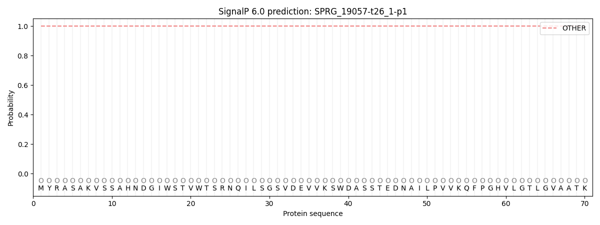
SignalP and Lipop Annotations help
This protein is predicted as OTHER
| Other | SP_Sec_SPI | CS Position |
|---|---|---|
| 0.999901 | 0.000138 |