You are browsing environment: FUNGIDB
CAZyme Information: SDRG_02625-t26_1-p1
You are here: Home > Sequence: SDRG_02625-t26_1-p1
Basic Information |
Genomic context |
Full Sequence |
Enzyme annotations |
CAZy signature domains |
CDD domains |
CAZyme hits |
PDB hits |
Swiss-Prot hits |
SignalP and Lipop annotations |
TMHMM annotations
Basic Information help
| Species | Saprolegnia diclina | |||||||||||
|---|---|---|---|---|---|---|---|---|---|---|---|---|
| Lineage | Oomycota; NA; ; Saprolegniaceae; Saprolegnia; Saprolegnia diclina | |||||||||||
| CAZyme ID | SDRG_02625-t26_1-p1 | |||||||||||
| CAZy Family | CBM9 | |||||||||||
| CAZyme Description | hypothetical protein | |||||||||||
| CAZyme Property |
|
|||||||||||
| Genome Property |
|
|||||||||||
| Gene Location | ||||||||||||
CAZyme Signature Domains help
| Family | Start | End | Evalue | family coverage |
|---|---|---|---|---|
| GH81 | 111 | 791 | 7.8e-147 | 0.9790996784565916 |
CDD Domains download full data without filtering help
| Cdd ID | Domain | E-Value | qStart | qEnd | sStart | sEnd | Domain Description |
|---|---|---|---|---|---|---|---|
| 407548 | Glyco_hydro81C | 3.13e-83 | 450 | 789 | 4 | 349 | Glycosyl hydrolase family 81 C-terminal domain. Family of eukaryotic beta-1,3-glucanases. Within the Aspergillus fumigatus protein, two perfectly conserved Glu residues (E550 or E554) have been proposed as putative nucleophiles of the active site of the Engl1 endoglucanase, while the proton donor would be D475. The endo-beta-1,3-glucanase activity is essential for efficient spore release. This entry represents the helical C-terminal domain. |
| 227785 | Acf2 | 8.85e-75 | 116 | 789 | 88 | 749 | Endoglucanase Acf2 [Carbohydrate transport and metabolism]. |
| 397619 | Glyco_hydro_81 | 6.45e-28 | 107 | 442 | 4 | 316 | Glycosyl hydrolase family 81. Family of eukaryotic beta-1,3-glucanases. Within the Aspergillus fumigatus protein ENGL1, two perfectly conserved Glu residues (E550 or E554) have been proposed as putative nucleophiles of the active site of the Engl1 endoglucanase, while the proton donor would be D475. The endo-beta-1,3-glucanase activity is essential for efficient spore release. |
| 176179 | enoyl_red | 0.008 | 241 | 416 | 41 | 204 | enoyl reductase of polyketide synthase. Putative enoyl reductase of polyketide synthase. Polyketide synthases produce polyketides in step by step mechanism that is similar to fatty acid synthesis. Enoyl reductase reduces a double to single bond. Erythromycin is one example of a polyketide generated by 3 complex enzymes (megasynthases). 2-enoyl thioester reductase (ETR) catalyzes the NADPH-dependent dependent conversion of trans-2-enoyl acyl carrier protein/coenzyme A (ACP/CoA) to acyl-(ACP/CoA) in fatty acid synthesis. 2-enoyl thioester reductase activity has been linked in Candida tropicalis as essential in maintaining mitiochondrial respiratory function. This ETR family is a part of the medium chain dehydrogenase/reductase family, but lack the zinc coordination sites characteristic of the alcohol dehydrogenases in this family. NAD(P)(H)-dependent oxidoreductases are the major enzymes in the interconversion of alcohols and aldehydes or ketones. Alcohol dehydrogenase in the liver converts ethanol and NAD+ to acetaldehyde and NADH, while in yeast and some other microorganisms ADH catalyzes the conversion acetaldehyde to ethanol in alcoholic fermentation. ADH is a member of the medium chain alcohol dehydrogenase family (MDR), which has a NAD(P)(H)-binding domain in a Rossmann fold of a beta-alpha form. The NAD(H)-binding region is comprised of 2 structurally similar halves, each of which contacts a mononucleotide. The N-terminal catalytic domain has a distant homology to GroES. These proteins typically form dimers (typically higher plants, mammals) or tetramers (yeast, bacteria), and have 2 tightly bound zinc atoms per subunit, a catalytic zinc at the active site, and a structural zinc in a lobe of the catalytic domain. NAD(H) binding occurs in the cleft between the catalytic and coenzyme-binding domains, at the active site, and coenzyme binding induces a conformational closing of this cleft. Coenzyme binding typically precedes and contributes to substrate binding. |
CAZyme Hits help
| Hit ID | E-Value | Query Start | Query End | Hit Start | Hit End |
|---|---|---|---|---|---|
| 1.44e-189 | 96 | 795 | 71 | 815 | |
| 5.11e-116 | 81 | 796 | 64 | 718 | |
| 6.56e-116 | 93 | 796 | 55 | 702 | |
| 3.40e-115 | 93 | 796 | 66 | 713 | |
| 1.96e-112 | 94 | 592 | 74 | 606 |
PDB Hits download full data without filtering help
| Hit ID | E-Value | Query Start | Query End | Hit Start | Hit End | Description |
|---|---|---|---|---|---|---|
| 5.57e-58 | 109 | 796 | 31 | 719 | The structure of a glycoside hydrolase family 81 endo-[beta]-1,3-glucanase [Rhizomucor miehei],4K3A_B The structure of a glycoside hydrolase family 81 endo-[beta]-1,3-glucanase [Rhizomucor miehei] |
|
| 7.51e-58 | 109 | 796 | 56 | 744 | Crystal structure of GH family 81 beta-1,3-glucanase from Rhizomucr miehei complexed with laminaripentaose [Rhizomucor miehei],5XBZ_B Crystal structure of GH family 81 beta-1,3-glucanase from Rhizomucr miehei complexed with laminaripentaose [Rhizomucor miehei],5XC2_A Crystal structure of GH family 81 beta-1,3-glucanase from Rhizomucr miehei complexed with laminarihexaose [Rhizomucor miehei],5XC2_B Crystal structure of GH family 81 beta-1,3-glucanase from Rhizomucr miehei complexed with laminarihexaose [Rhizomucor miehei],5XC2_C Crystal structure of GH family 81 beta-1,3-glucanase from Rhizomucr miehei complexed with laminarihexaose [Rhizomucor miehei],5XC2_D Crystal structure of GH family 81 beta-1,3-glucanase from Rhizomucr miehei complexed with laminarihexaose [Rhizomucor miehei] |
|
| 1.66e-56 | 110 | 796 | 32 | 719 | The structure of a glycoside hydrolase family 81 endo-[beta]-1,3-glucanase [Rhizomucor miehei],4K35_B The structure of a glycoside hydrolase family 81 endo-[beta]-1,3-glucanase [Rhizomucor miehei] |
|
| 1.57e-35 | 414 | 792 | 279 | 653 | Chain A, Glycoside hydrolase family 81 [Acetivibrio thermocellus ATCC 27405] |
|
| 6.19e-25 | 403 | 792 | 291 | 689 | Chain A, Glycoside Hydrolase [Halalkalibacterium halodurans C-125] |
Swiss-Prot Hits download full data without filtering help
| Hit ID | E-Value | Query Start | Query End | Hit Start | Hit End | Description |
|---|---|---|---|---|---|---|
| 3.37e-51 | 80 | 793 | 179 | 900 | Probable endo-1,3(4)-beta-glucanase ARB_01444 OS=Arthroderma benhamiae (strain ATCC MYA-4681 / CBS 112371) OX=663331 GN=ARB_01444 PE=1 SV=1 |
|
| 1.86e-48 | 155 | 793 | 117 | 735 | Primary septum endo-1,3(4)-beta-glucanase OS=Schizosaccharomyces pombe (strain 972 / ATCC 24843) OX=284812 GN=eng1 PE=3 SV=1 |
|
| 7.99e-43 | 118 | 793 | 35 | 700 | Ascus wall endo-1,3(4)-beta-glucanase OS=Schizosaccharomyces pombe (strain 972 / ATCC 24843) OX=284812 GN=eng2 PE=1 SV=1 |
|
| 4.62e-39 | 116 | 789 | 432 | 1106 | Endo-1,3(4)-beta-glucanase 1 OS=Saccharomyces cerevisiae (strain ATCC 204508 / S288c) OX=559292 GN=DSE4 PE=1 SV=1 |
|
| 2.01e-37 | 372 | 771 | 355 | 750 | Endo-1,3(4)-beta-glucanase 2 OS=Saccharomyces cerevisiae (strain ATCC 204508 / S288c) OX=559292 GN=ACF2 PE=1 SV=1 |
SignalP and Lipop Annotations help
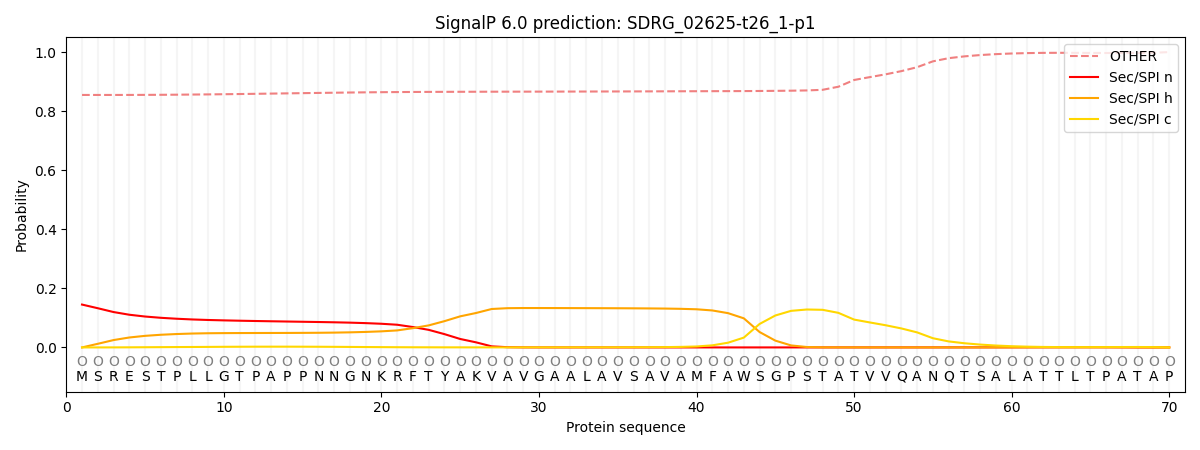
This protein is predicted as OTHER

| Other | SP_Sec_SPI | CS Position |
|---|---|---|
| 0.861217 | 0.138783 |
