You are browsing environment: FUNGIDB
CAZyme Information: SAPIO_CDS10803-t41_1-p1
You are here: Home > Sequence: SAPIO_CDS10803-t41_1-p1
Basic Information |
Genomic context |
Full Sequence |
Enzyme annotations |
CAZy signature domains |
CDD domains |
CAZyme hits |
PDB hits |
Swiss-Prot hits |
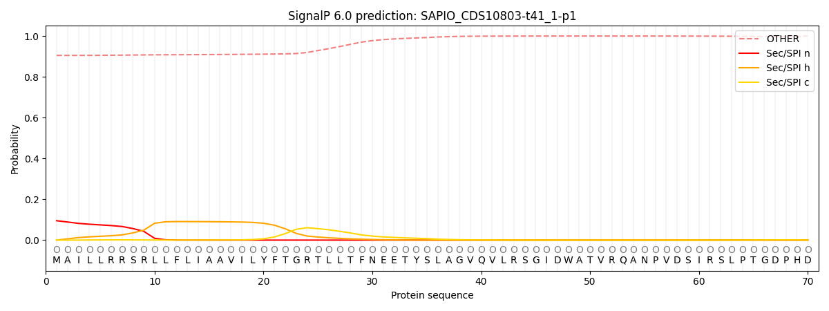
SignalP and Lipop annotations |
TMHMM annotations
Basic Information help
| Species | Scedosporium apiospermum | |||||||||||
|---|---|---|---|---|---|---|---|---|---|---|---|---|
| Lineage | Ascomycota; Sordariomycetes; ; Microascaceae; Scedosporium; Scedosporium apiospermum | |||||||||||
| CAZyme ID | SAPIO_CDS10803-t41_1-p1 | |||||||||||
| CAZy Family | AA7 | |||||||||||
| CAZyme Description | hypothetical protein | |||||||||||
| CAZyme Property |
|
|||||||||||
| Genome Property |
|
|||||||||||
| Gene Location | ||||||||||||
Enzyme Prediction help
| EC | 3.2.1.113:7 |
|---|
CAZyme Signature Domains help
| Family | Start | End | Evalue | family coverage |
|---|---|---|---|---|
| GH47 | 102 | 572 | 2.4e-161 | 0.9955156950672646 |
CDD Domains download full data without filtering help
| Cdd ID | Domain | E-Value | qStart | qEnd | sStart | sEnd | Domain Description |
|---|---|---|---|---|---|---|---|
| 396217 | Glyco_hydro_47 | 0.0 | 102 | 572 | 2 | 453 | Glycosyl hydrolase family 47. Members of this family are alpha-mannosidases that catalyze the hydrolysis of the terminal 1,2-linked alpha-D-mannose residues in the oligo-mannose oligosaccharide Man(9)(GlcNAc)(2). |
| 240427 | PTZ00470 | 4.17e-126 | 92 | 575 | 70 | 521 | glycoside hydrolase family 47 protein; Provisional |
| 395844 | Aminotran_4 | 1.02e-14 | 634 | 856 | 14 | 216 | Amino-transferase class IV. The D-amino acid transferases (D-AAT) are required by bacteria to catalyze the synthesis of D-glutamic acid and D-alanine, which are essential constituents of bacterial cell wall and are the building block for other D-amino acids. Despite the difference in the structure of the substrates, D-AATs and L-ATTs have strong similarity. |
| 169002 | PRK07546 | 1.97e-13 | 652 | 860 | 12 | 208 | hypothetical protein; Provisional |
| 238800 | ADCL_like | 5.03e-05 | 772 | 851 | 145 | 221 | ADCL_like: 4-Amino-4-deoxychorismate lyase: is a member of the fold-type IV of PLP dependent enzymes that converts 4-amino-4-deoxychorismate (ADC) to p-aminobenzoate and pyruvate. Based on the information available from the crystal structure, most members of this subgroup are likely to function as dimers. The enzyme from E.Coli, the structure of which is available, is a homodimer that is folded into a small and a larger domain. The coenzyme pyridoxal 5; -phosphate resides at the interface of the two domains that is linked by a flexible loop. Members of this subgroup are found in Eukaryotes and bacteria. |
CAZyme Hits help
| Hit ID | E-Value | Query Start | Query End | Hit Start | Hit End |
|---|---|---|---|---|---|
| 8.20e-210 | 44 | 578 | 53 | 575 | |
| 5.76e-207 | 30 | 574 | 34 | 588 | |
| 4.80e-206 | 42 | 578 | 51 | 575 | |
| 2.51e-204 | 44 | 574 | 45 | 569 | |
| 1.86e-200 | 46 | 582 | 53 | 575 |
PDB Hits download full data without filtering help
| Hit ID | E-Value | Query Start | Query End | Hit Start | Hit End | Description |
|---|---|---|---|---|---|---|
| 2.85e-73 | 92 | 569 | 3 | 470 | Penicillium citrinum alpha-1,2-mannosidase complex with glycerol [Penicillium citrinum],2RI8_B Penicillium citrinum alpha-1,2-mannosidase complex with glycerol [Penicillium citrinum],2RI9_A Penicillium citrinum alpha-1,2-mannosidase in complex with a substrate analog [Penicillium citrinum],2RI9_B Penicillium citrinum alpha-1,2-mannosidase in complex with a substrate analog [Penicillium citrinum] |
|
| 7.20e-73 | 92 | 569 | 38 | 505 | Structure of P. citrinum alpha 1,2-mannosidase reveals the basis for differences in specificity of the ER and Golgi Class I enzymes [Penicillium citrinum],1KKT_B Structure of P. citrinum alpha 1,2-mannosidase reveals the basis for differences in specificity of the ER and Golgi Class I enzymes [Penicillium citrinum],1KRE_A Structure Of P. Citrinum Alpha 1,2-Mannosidase Reveals The Basis For Differences In Specificity Of The Er And Golgi Class I Enzymes [Penicillium citrinum],1KRE_B Structure Of P. Citrinum Alpha 1,2-Mannosidase Reveals The Basis For Differences In Specificity Of The Er And Golgi Class I Enzymes [Penicillium citrinum],1KRF_A Structure Of P. Citrinum Alpha 1,2-mannosidase Reveals The Basis For Differences In Specificity Of The Er And Golgi Class I Enzymes [Penicillium citrinum],1KRF_B Structure Of P. Citrinum Alpha 1,2-mannosidase Reveals The Basis For Differences In Specificity Of The Er And Golgi Class I Enzymes [Penicillium citrinum] |
|
| 4.97e-67 | 91 | 569 | 2 | 448 | Crystal structure of the class I human endoplasmic reticulum 1,2-alpha-mannosidase and Man9GlcNAc2-PA complex [Homo sapiens] |
|
| 5.80e-67 | 91 | 569 | 7 | 453 | Crystal Structure Of Human Class I Alpha1,2-Mannosidase [Homo sapiens] |
|
| 5.85e-67 | 49 | 569 | 24 | 531 | Crystal Structure Of Human Class I alpha-1,2-Mannosidase In Complex With Thio-Disaccharide Substrate Analogue [Homo sapiens] |
Swiss-Prot Hits download full data without filtering help
| Hit ID | E-Value | Query Start | Query End | Hit Start | Hit End | Description |
|---|---|---|---|---|---|---|
| 9.60e-75 | 92 | 571 | 37 | 506 | Probable mannosyl-oligosaccharide alpha-1,2-mannosidase 1B OS=Aspergillus terreus (strain NIH 2624 / FGSC A1156) OX=341663 GN=mns1B PE=3 SV=1 |
|
| 1.76e-74 | 83 | 571 | 31 | 513 | Mannosyl-oligosaccharide alpha-1,2-mannosidase OS=Coccidioides posadasii (strain RMSCC 757 / Silveira) OX=443226 GN=CPSG_02648 PE=1 SV=1 |
|
| 1.08e-73 | 87 | 571 | 35 | 509 | Probable mannosyl-oligosaccharide alpha-1,2-mannosidase 1B OS=Aspergillus niger (strain CBS 513.88 / FGSC A1513) OX=425011 GN=mns1B PE=3 SV=1 |
|
| 1.49e-73 | 87 | 571 | 35 | 509 | Mannosyl-oligosaccharide alpha-1,2-mannosidase 1B OS=Aspergillus phoenicis OX=5063 GN=mns1B PE=2 SV=1 |
|
| 3.70e-72 | 92 | 569 | 38 | 505 | Mannosyl-oligosaccharide alpha-1,2-mannosidase OS=Penicillium citrinum OX=5077 GN=MSDC PE=1 SV=2 |
SignalP and Lipop Annotations help
This protein is predicted as OTHER

| Other | SP_Sec_SPI | CS Position |
|---|---|---|
| 0.910137 | 0.089884 |
