You are browsing environment: FUNGIDB
CAZyme Information: RVD83193.1
You are here: Home > Sequence: RVD83193.1
Basic Information |
Genomic context |
Full Sequence |
Enzyme annotations |
CAZy signature domains |
CDD domains |
CAZyme hits |
PDB hits |
Swiss-Prot hits |
SignalP and Lipop annotations |
TMHMM annotations
Basic Information help
| Species | Arthrobotrys flagrans | |||||||||||
|---|---|---|---|---|---|---|---|---|---|---|---|---|
| Lineage | Ascomycota; Orbiliomycetes; ; Orbiliaceae; Arthrobotrys; Arthrobotrys flagrans | |||||||||||
| CAZyme ID | RVD83193.1 | |||||||||||
| CAZy Family | CE5 | |||||||||||
| CAZyme Description | unspecified product | |||||||||||
| CAZyme Property |
|
|||||||||||
| Genome Property |
|
|||||||||||
| Gene Location | ||||||||||||
CAZyme Signature Domains help
| Family | Start | End | Evalue | family coverage |
|---|---|---|---|---|
| GH5 | 542 | 827 | 3.6e-105 | 0.9931740614334471 |
CDD Domains download full data without filtering help
| Cdd ID | Domain | E-Value | qStart | qEnd | sStart | sEnd | Domain Description |
|---|---|---|---|---|---|---|---|
| 225184 | CitE | 1.22e-70 | 85 | 391 | 1 | 272 | Citrate lyase beta subunit [Carbohydrate transport and metabolism]. |
| 397422 | HpcH_HpaI | 2.15e-30 | 84 | 336 | 1 | 221 | HpcH/HpaI aldolase/citrate lyase family. This family includes 2,4-dihydroxyhept-2-ene-1,7-dioic acid aldolase and 4-hydroxy-2-oxovalerate aldolase. |
| 225344 | BglC | 1.93e-19 | 488 | 830 | 13 | 322 | Aryl-phospho-beta-D-glucosidase BglC, GH1 family [Carbohydrate transport and metabolism]. |
| 406131 | C-C_Bond_Lyase | 1.23e-12 | 87 | 393 | 11 | 314 | C-C_Bond_Lyase of the TIM-Barrel fold. This family of TIM-Barrel fold C-C bond lyase is related to citrate-lyase. These genes are found in the biosynthetic operon, with other enzymatic domains, associated with the Ter stress response operon and are predicted to be involved in the biosynthesis of a ribo-nucleoside involved in stress response. |
CAZyme Hits help
| Hit ID | E-Value | Query Start | Query End | Hit Start | Hit End |
|---|---|---|---|---|---|
| 0.0 | 439 | 979 | 4 | 544 | |
| 2.22e-145 | 457 | 977 | 16 | 530 | |
| 3.69e-144 | 469 | 977 | 27 | 543 | |
| 1.74e-136 | 479 | 976 | 50 | 540 | |
| 5.83e-134 | 479 | 977 | 52 | 542 |
PDB Hits download full data without filtering help
| Hit ID | E-Value | Query Start | Query End | Hit Start | Hit End | Description |
|---|---|---|---|---|---|---|
| 1.54e-41 | 83 | 399 | 22 | 316 | Crystal Structure Analysis of human CLYBL in complex with free CoASH [Homo sapiens],5VXO_A Crystal Structure Analysis of human CLYBL in complex with propionyl-CoA [Homo sapiens],5VXO_B Crystal Structure Analysis of human CLYBL in complex with propionyl-CoA [Homo sapiens],5VXO_C Crystal Structure Analysis of human CLYBL in complex with propionyl-CoA [Homo sapiens],5VXS_A Crystal Structure Analysis of human CLYBL in apo form [Homo sapiens],5VXS_B Crystal Structure Analysis of human CLYBL in apo form [Homo sapiens],5VXS_C Crystal Structure Analysis of human CLYBL in apo form [Homo sapiens],5VXS_D Crystal Structure Analysis of human CLYBL in apo form [Homo sapiens],5VXS_E Crystal Structure Analysis of human CLYBL in apo form [Homo sapiens],5VXS_F Crystal Structure Analysis of human CLYBL in apo form [Homo sapiens] |
|
| 4.81e-29 | 81 | 393 | 12 | 317 | Malyl-CoA lyase from Methylobacterium extorquens [Methylorubrum extorquens AM1] |
|
| 1.83e-25 | 80 | 397 | 32 | 337 | Chain A, Malyl-CoA lyase [Cereibacter sphaeroides 2.4.1],4L9Y_B Chain B, Malyl-CoA lyase [Cereibacter sphaeroides 2.4.1],4L9Y_C Chain C, Malyl-CoA lyase [Cereibacter sphaeroides 2.4.1],4L9Y_D Chain D, Malyl-CoA lyase [Cereibacter sphaeroides 2.4.1],4L9Y_E Chain E, Malyl-CoA lyase [Cereibacter sphaeroides 2.4.1],4L9Y_F Chain F, Malyl-CoA lyase [Cereibacter sphaeroides 2.4.1],4L9Z_A Chain A, Malyl-CoA lyase [Cereibacter sphaeroides 2.4.1],4L9Z_B Chain B, Malyl-CoA lyase [Cereibacter sphaeroides 2.4.1],4L9Z_C Chain C, Malyl-CoA lyase [Cereibacter sphaeroides 2.4.1],4L9Z_D Chain D, Malyl-CoA lyase [Cereibacter sphaeroides 2.4.1],4L9Z_E Chain E, Malyl-CoA lyase [Cereibacter sphaeroides 2.4.1],4L9Z_F Chain F, Malyl-CoA lyase [Cereibacter sphaeroides 2.4.1] |
|
| 3.63e-23 | 82 | 392 | 7 | 281 | Crystal structure of citrate lyase beta subunit [Deinococcus radiodurans],1SGJ_B Crystal structure of citrate lyase beta subunit [Deinococcus radiodurans],1SGJ_C Crystal structure of citrate lyase beta subunit [Deinococcus radiodurans] |
|
| 2.21e-22 | 84 | 393 | 46 | 311 | Crystal Structure of RipC from Yersinia pestis [Yersinia pestis],3QLL_B Crystal Structure of RipC from Yersinia pestis [Yersinia pestis],3QLL_C Crystal Structure of RipC from Yersinia pestis [Yersinia pestis] |
Swiss-Prot Hits download full data without filtering help
| Hit ID | E-Value | Query Start | Query End | Hit Start | Hit End | Description |
|---|---|---|---|---|---|---|
| 4.43e-66 | 484 | 910 | 5 | 415 | Glucan 1,3-beta-glucosidase 3 OS=Schizosaccharomyces pombe (strain 972 / ATCC 24843) OX=284812 GN=exg3 PE=3 SV=1 |
|
| 1.27e-57 | 480 | 972 | 32 | 498 | Uncharacterized glycosyl hydrolase YBR056W OS=Saccharomyces cerevisiae (strain ATCC 204508 / S288c) OX=559292 GN=YBR056W PE=1 SV=1 |
|
| 1.74e-41 | 83 | 399 | 43 | 337 | Citramalyl-CoA lyase, mitochondrial OS=Mus musculus OX=10090 GN=Clybl PE=1 SV=2 |
|
| 8.05e-41 | 83 | 399 | 43 | 337 | Citramalyl-CoA lyase, mitochondrial OS=Rattus norvegicus OX=10116 GN=Clybl PE=2 SV=2 |
|
| 1.15e-40 | 83 | 399 | 45 | 339 | Citramalyl-CoA lyase, mitochondrial OS=Homo sapiens OX=9606 GN=CLYBL PE=1 SV=2 |
SignalP and Lipop Annotations help
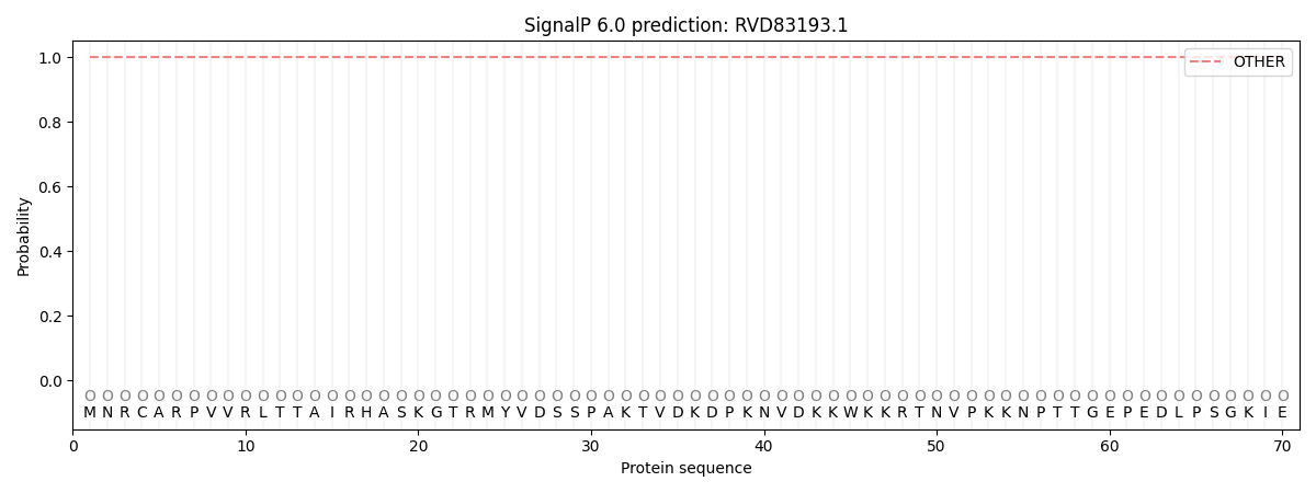
This protein is predicted as OTHER

| Other | SP_Sec_SPI | CS Position |
|---|---|---|
| 1.000063 | 0.000000 |
