You are browsing environment: FUNGIDB
CAZyme Information: RL4_JR_03201-RA-p1
You are here: Home > Sequence: RL4_JR_03201-RA-p1
Basic Information |
Genomic context |
Full Sequence |
Enzyme annotations |
CAZy signature domains |
CDD domains |
CAZyme hits |
PDB hits |
Swiss-Prot hits |
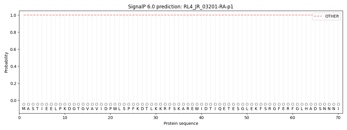
SignalP and Lipop annotations |
TMHMM annotations
Basic Information help
| Species | Raffaelea lauricola | |||||||||||
|---|---|---|---|---|---|---|---|---|---|---|---|---|
| Lineage | Ascomycota; Sordariomycetes; ; Ophiostomataceae; Raffaelea; Raffaelea lauricola | |||||||||||
| CAZyme ID | RL4_JR_03201-RA-p1 | |||||||||||
| CAZy Family | GH10 | |||||||||||
| CAZyme Description | unspecified product | |||||||||||
| CAZyme Property |
|
|||||||||||
| Genome Property |
|
|||||||||||
| Gene Location | ||||||||||||
Enzyme Prediction help
| EC | 2.4.1.18:166 | 2.4.1.18:50 |
|---|
CAZyme Signature Domains help
| Family | Start | End | Evalue | family coverage |
|---|---|---|---|---|
| GH13 | 235 | 528 | 2e-153 | 0.9965870307167235 |
CDD Domains download full data without filtering help
| Cdd ID | Domain | E-Value | qStart | qEnd | sStart | sEnd | Domain Description |
|---|---|---|---|---|---|---|---|
| 200460 | AmyAc_bac_euk_BE | 0.0 | 169 | 572 | 3 | 406 | Alpha amylase catalytic domain found in bacterial and eukaryotic branching enzymes. Branching enzymes (BEs) catalyze the formation of alpha-1,6 branch points in either glycogen or starch by cleavage of the alpha-1,4 glucosidic linkage yielding a non-reducing end oligosaccharide chain, and subsequent attachment to the alpha-1,6 position. By increasing the number of non-reducing ends, glycogen is more reactive to synthesis and digestion as well as being more soluble. This group includes bacterial and eukaryotic proteins. The Alpha-amylase family comprises the largest family of glycoside hydrolases (GH), with the majority of enzymes acting on starch, glycogen, and related oligo- and polysaccharides. These proteins catalyze the transformation of alpha-1,4 and alpha-1,6 glucosidic linkages with retention of the anomeric center. The protein is described as having 3 domains: A, B, C. A is a (beta/alpha) 8-barrel; B is a loop between the beta 3 strand and alpha 3 helix of A; C is the C-terminal extension characterized by a Greek key. The majority of the enzymes have an active site cleft found between domains A and B where a triad of catalytic residues (Asp, Glu and Asp) performs catalysis. Other members of this family have lost the catalytic activity as in the case of the human 4F2hc, or only have 2 residues that serve as the catalytic nucleophile and the acid/base, such as Thermus A4 beta-galactosidase with 2 Glu residues (GH42) and human alpha-galactosidase with 2 Asp residues (GH31). The family members are quite extensive and include: alpha amylase, maltosyltransferase, cyclodextrin glycotransferase, maltogenic amylase, neopullulanase, isoamylase, 1,4-alpha-D-glucan maltotetrahydrolase, 4-alpha-glucotransferase, oligo-1,6-glucosidase, amylosucrase, sucrose phosphorylase, and amylomaltase. |
| 214349 | atpB | 0.0 | 706 | 1171 | 17 | 489 | ATP synthase CF1 beta subunit |
| 211621 | atpD | 0.0 | 706 | 1168 | 3 | 461 | ATP synthase, F1 beta subunit. The sequences of ATP synthase F1 alpha and beta subunits are related and both contain a nucleotide-binding site for ATP and ADP. They have a common amino terminal domain but vary at the C-terminus. The beta chain has catalytic activity, while the alpha chain is a regulatory subunit. Proton translocating ATP synthase, F1 beta subunit is homologous to proton translocating ATP synthase archaeal/vacuolar(V1), A subunit. [Energy metabolism, ATP-proton motive force interconversion] |
| 215246 | PLN02447 | 0.0 | 9 | 663 | 56 | 709 | 1,4-alpha-glucan-branching enzyme |
| 410877 | F1-ATPase_beta_CD | 0.0 | 779 | 1055 | 1 | 277 | F1 ATP synthase beta subunit, central domain. The F-ATPase is found in bacterial plasma membranes, mitochondrial inner membranes and in chloroplast thylakoid membranes. It has also been found in the archaea Methanosarcina barkeri. It uses a proton gradient to drive ATP synthesis and hydrolyzes ATP to build the proton gradient. The mitochondrial extrinsic membrane domain, F1, is composed of alpha, beta, gamma, delta and epsilon subunits with a stoichiometry of 3:3:1:1:1. The beta subunit of ATP synthase is catalytic. Alpha and beta subunits form the globular catalytic moiety, a hexameric ring of alternating alpha and beta subunits. Gamma, delta and epsilon subunits form a stalk, connecting F1 to F0, the integral membrane proton-translocating domain. |
CAZyme Hits help
| Hit ID | E-Value | Query Start | Query End | Hit Start | Hit End |
|---|---|---|---|---|---|
| 0.0 | 8 | 659 | 24 | 674 | |
| 0.0 | 7 | 659 | 22 | 674 | |
| 0.0 | 7 | 660 | 23 | 675 | |
| 0.0 | 7 | 660 | 23 | 675 | |
| 0.0 | 7 | 660 | 23 | 675 |
PDB Hits download full data without filtering help
| Hit ID | E-Value | Query Start | Query End | Hit Start | Hit End | Description |
|---|---|---|---|---|---|---|
| 9.99e-311 | 18 | 658 | 34 | 671 | Crystal structure of human glycogen branching enzyme (GBE1) [Homo sapiens],4BZY_B Crystal structure of human glycogen branching enzyme (GBE1) [Homo sapiens],4BZY_C Crystal structure of human glycogen branching enzyme (GBE1) [Homo sapiens] |
|
| 2.15e-308 | 22 | 658 | 6 | 639 | Crystal structure of human glycogen branching enzyme (GBE1) in complex with acarbose [Homo sapiens],5CLT_B Crystal structure of human glycogen branching enzyme (GBE1) in complex with acarbose [Homo sapiens],5CLT_C Crystal structure of human glycogen branching enzyme (GBE1) in complex with acarbose [Homo sapiens],5CLW_A Crystal structure of human glycogen branching enzyme (GBE1) in complex with maltoheptaose [Homo sapiens],5CLW_B Crystal structure of human glycogen branching enzyme (GBE1) in complex with maltoheptaose [Homo sapiens],5CLW_C Crystal structure of human glycogen branching enzyme (GBE1) in complex with maltoheptaose [Homo sapiens] |
|
| 3.42e-265 | 1 | 660 | 1 | 660 | Structure of the Starch Branching Enzyme I (BEI) from Oryza sativa L [Oryza sativa Japonica Group] |
|
| 5.52e-265 | 699 | 1171 | 1 | 474 | Chain D, ATP synthase beta subunit [Ogataea angusta],5LQX_E Chain E, ATP synthase beta subunit [Ogataea angusta],5LQX_F Chain F, ATP synthase beta subunit [Ogataea angusta],5LQY_D Chain D, ATP synthase beta subunit [Ogataea angusta],5LQY_E Chain E, ATP synthase beta subunit [Ogataea angusta],5LQY_F Chain F, ATP synthase beta subunit [Ogataea angusta],5LQZ_D Chain D, ATP synthase beta subunit [Ogataea angusta],5LQZ_E Chain E, ATP synthase beta subunit [Ogataea angusta],5LQZ_F Chain F, ATP synthase beta subunit [Ogataea angusta] |
|
| 9.67e-265 | 1 | 660 | 1 | 660 | Structure of the Starch Branching Enzyme I (BEI) complexed with maltopentaose from Oryza sativa L [Oryza sativa Japonica Group],3VU2_B Structure of the Starch Branching Enzyme I (BEI) complexed with maltopentaose from Oryza sativa L [Oryza sativa Japonica Group] |
Swiss-Prot Hits download full data without filtering help
| Hit ID | E-Value | Query Start | Query End | Hit Start | Hit End | Description |
|---|---|---|---|---|---|---|
| 0.0 | 661 | 1173 | 7 | 516 | ATP synthase subunit beta, mitochondrial OS=Neurospora crassa (strain ATCC 24698 / 74-OR23-1A / CBS 708.71 / DSM 1257 / FGSC 987) OX=367110 GN=atp-2 PE=2 SV=1 |
|
| 0.0 | 14 | 660 | 15 | 658 | 1,4-alpha-glucan-branching enzyme OS=Rhizophagus irregularis (strain DAOM 181602 / DAOM 197198 / MUCL 43194) OX=747089 GN=GLC3 PE=2 SV=2 |
|
| 0.0 | 19 | 660 | 9 | 660 | 1,4-alpha-glucan-branching enzyme OS=Yarrowia lipolytica (strain CLIB 122 / E 150) OX=284591 GN=GLC3 PE=3 SV=1 |
|
| 0.0 | 2 | 663 | 4 | 664 | 1,4-alpha-glucan-branching enzyme OS=Aspergillus oryzae (strain ATCC 42149 / RIB 40) OX=510516 GN=gbeA PE=2 SV=1 |
|
| 0.0 | 11 | 660 | 8 | 658 | 1,4-alpha-glucan-branching enzyme OS=Cryptococcus neoformans var. neoformans serotype D (strain JEC21 / ATCC MYA-565) OX=214684 GN=GLC3 PE=3 SV=1 |
SignalP and Lipop Annotations help
This protein is predicted as OTHER

| Other | SP_Sec_SPI | CS Position |
|---|---|---|
| 1.000046 | 0.000000 |
