You are browsing environment: FUNGIDB
CAZyme Information: PWY71963.1
You are here: Home > Sequence: PWY71963.1
Basic Information |
Genomic context |
Full Sequence |
Enzyme annotations |
CAZy signature domains |
CDD domains |
CAZyme hits |
PDB hits |
Swiss-Prot hits |
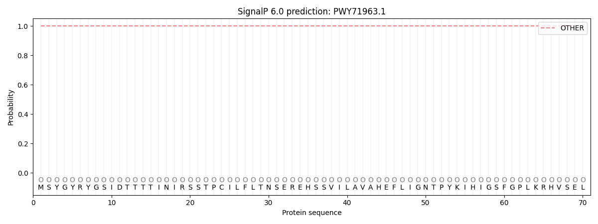
SignalP and Lipop annotations |
TMHMM annotations
Basic Information help
| Species | Aspergillus eucalypticola | |||||||||||
|---|---|---|---|---|---|---|---|---|---|---|---|---|
| Lineage | Ascomycota; Eurotiomycetes; ; Aspergillaceae; Aspergillus; Aspergillus eucalypticola | |||||||||||
| CAZyme ID | PWY71963.1 | |||||||||||
| CAZy Family | GT90 | |||||||||||
| CAZyme Description | UDP-Glycosyltransferase/glycogen phosphorylase | |||||||||||
| CAZyme Property |
|
|||||||||||
| Genome Property |
|
|||||||||||
| Gene Location | ||||||||||||
CDD Domains download full data without filtering help
| Cdd ID | Domain | E-Value | qStart | qEnd | sStart | sEnd | Domain Description |
|---|---|---|---|---|---|---|---|
| 224995 | MmsB | 2.00e-46 | 465 | 741 | 14 | 282 | 3-hydroxyisobutyrate dehydrogenase or related beta-hydroxyacid dehydrogenase [Lipid transport and metabolism]. |
| 397486 | NAD_binding_2 | 1.37e-27 | 465 | 606 | 13 | 149 | NAD binding domain of 6-phosphogluconate dehydrogenase. The NAD binding domain of 6-phosphogluconate dehydrogenase adopts a Rossmann fold. |
| 340817 | GT1_Gtf-like | 4.16e-18 | 105 | 444 | 54 | 386 | UDP-glycosyltransferases and similar proteins. This family includes the Gtfs, a group of homologous glycosyltransferases involved in the final stages of the biosynthesis of antibiotics vancomycin and related chloroeremomycin. Gtfs transfer sugar moieties from an activated NDP-sugar donor to the oxidatively cross-linked heptapeptide core of vancomycin group antibiotics. The core structure is important for the bioactivity of the antibiotics. |
| 183197 | garR | 1.00e-16 | 466 | 714 | 17 | 256 | tartronate semialdehyde reductase; Provisional |
| 185358 | PRK15461 | 2.24e-16 | 466 | 751 | 16 | 293 | sulfolactaldehyde 3-reductase. |
CAZyme Hits help
| Hit ID | E-Value | Query Start | Query End | Hit Start | Hit End |
|---|---|---|---|---|---|
| 0.0 | 1 | 467 | 1 | 499 | |
| 0.0 | 1 | 467 | 1 | 499 | |
| 3.02e-284 | 1 | 467 | 1 | 497 | |
| 1.67e-211 | 23 | 754 | 4 | 829 | |
| 5.44e-97 | 23 | 464 | 2 | 483 |
PDB Hits download full data without filtering help
| Hit ID | E-Value | Query Start | Query End | Hit Start | Hit End | Description |
|---|---|---|---|---|---|---|
| 2.87e-15 | 480 | 750 | 31 | 295 | Chain A, 6-phosphogluconate dehydrogenase NAD-binding [Dyadobacter fermentans DSM 18053],4GBJ_B Chain B, 6-phosphogluconate dehydrogenase NAD-binding [Dyadobacter fermentans DSM 18053],4GBJ_C Chain C, 6-phosphogluconate dehydrogenase NAD-binding [Dyadobacter fermentans DSM 18053],4GBJ_D Chain D, 6-phosphogluconate dehydrogenase NAD-binding [Dyadobacter fermentans DSM 18053] |
|
| 5.61e-10 | 526 | 714 | 90 | 275 | Structure of Glyoxylate reductase 1 from Arabidopsis (AtGLYR1) [Arabidopsis thaliana] |
|
| 7.01e-10 | 517 | 747 | 66 | 296 | The crystal structure of Bacillus cereus 3-hydroxyisobutyrate dehydrogenase in complex with NAD [Bacillus cereus ATCC 14579],5JE8_B The crystal structure of Bacillus cereus 3-hydroxyisobutyrate dehydrogenase in complex with NAD [Bacillus cereus ATCC 14579],5JE8_C The crystal structure of Bacillus cereus 3-hydroxyisobutyrate dehydrogenase in complex with NAD [Bacillus cereus ATCC 14579],5JE8_D The crystal structure of Bacillus cereus 3-hydroxyisobutyrate dehydrogenase in complex with NAD [Bacillus cereus ATCC 14579] |
|
| 9.29e-10 | 515 | 714 | 61 | 259 | Structural and Kinetic Properties of a beta-hydroxyacid dehydrogenase involved in nicotinate fermentation [Eubacterium barkeri],3CKY_B Structural and Kinetic Properties of a beta-hydroxyacid dehydrogenase involved in nicotinate fermentation [Eubacterium barkeri],3CKY_C Structural and Kinetic Properties of a beta-hydroxyacid dehydrogenase involved in nicotinate fermentation [Eubacterium barkeri],3CKY_D Structural and Kinetic Properties of a beta-hydroxyacid dehydrogenase involved in nicotinate fermentation [Eubacterium barkeri] |
|
| 9.69e-10 | 529 | 750 | 79 | 292 | Crystal structure of SLA Reductase YihU from E. Coli [Escherichia coli],6SM7_B Crystal structure of SLA Reductase YihU from E. Coli [Escherichia coli],6SM7_C Crystal structure of SLA Reductase YihU from E. Coli [Escherichia coli],6SM7_D Crystal structure of SLA Reductase YihU from E. Coli [Escherichia coli],6SMY_A Crystal structure of SLA Reductase YihU from E. Coli with NADH and product DHPS [Escherichia coli K-12],6SMY_B Crystal structure of SLA Reductase YihU from E. Coli with NADH and product DHPS [Escherichia coli K-12],6SMY_C Crystal structure of SLA Reductase YihU from E. Coli with NADH and product DHPS [Escherichia coli K-12],6SMY_D Crystal structure of SLA Reductase YihU from E. Coli with NADH and product DHPS [Escherichia coli K-12],6SMZ_A Crystal structure of SLA Reductase YihU from E. Coli in complex with NADH [Escherichia coli K-12],6SMZ_B Crystal structure of SLA Reductase YihU from E. Coli in complex with NADH [Escherichia coli K-12],6SMZ_C Crystal structure of SLA Reductase YihU from E. Coli in complex with NADH [Escherichia coli K-12],6SMZ_D Crystal structure of SLA Reductase YihU from E. Coli in complex with NADH [Escherichia coli K-12] |
Swiss-Prot Hits download full data without filtering help
| Hit ID | E-Value | Query Start | Query End | Hit Start | Hit End | Description |
|---|---|---|---|---|---|---|
| 2.48e-55 | 23 | 427 | 6 | 436 | Glycosyltransferase buaB OS=Aspergillus burnettii OX=2508778 GN=buaB PE=3 SV=1 |
|
| 1.44e-47 | 23 | 429 | 6 | 466 | Glycosyltransferase sdnJ OS=Sordaria araneosa OX=573841 GN=sdnJ PE=1 SV=1 |
|
| 3.79e-11 | 456 | 723 | 5 | 264 | Uncharacterized oxidoreductase YfjR OS=Bacillus subtilis (strain 168) OX=224308 GN=yfjR PE=3 SV=2 |
|
| 2.39e-09 | 526 | 714 | 69 | 254 | Glyoxylate/succinic semialdehyde reductase 1 OS=Arabidopsis thaliana OX=3702 GN=GLYR1 PE=1 SV=1 |
|
| 3.40e-09 | 414 | 653 | 136 | 371 | Putative oxidoreductase GLYR1 OS=Danio rerio OX=7955 GN=glyr1 PE=2 SV=1 |
SignalP and Lipop Annotations help
This protein is predicted as OTHER

| Other | SP_Sec_SPI | CS Position |
|---|---|---|
| 1.000045 | 0.000001 |
