You are browsing environment: FUNGIDB
CAZyme Information: PSK37157.1
You are here: Home > Sequence: PSK37157.1
Basic Information |
Genomic context |
Full Sequence |
Enzyme annotations |
CAZy signature domains |
CDD domains |
CAZyme hits |
PDB hits |
Swiss-Prot hits |
SignalP and Lipop annotations |
TMHMM annotations
Basic Information help
| Species | [Candida] pseudohaemulonis | |||||||||||
|---|---|---|---|---|---|---|---|---|---|---|---|---|
| Lineage | Ascomycota; Saccharomycetes; ; Debaryomycetaceae; Candida; [Candida] pseudohaemulonis | |||||||||||
| CAZyme ID | PSK37157.1 | |||||||||||
| CAZy Family | GH17 | |||||||||||
| CAZyme Description | unspecified product | |||||||||||
| CAZyme Property |
|
|||||||||||
| Genome Property |
|
|||||||||||
| Gene Location | ||||||||||||
Enzyme Prediction help
| EC | 2.4.1.186:3 |
|---|
CAZyme Signature Domains help
| Family | Start | End | Evalue | family coverage |
|---|---|---|---|---|
| GT8 | 7 | 229 | 2.9e-37 | 0.8638132295719845 |
CDD Domains download full data without filtering help
| Cdd ID | Domain | E-Value | qStart | qEnd | sStart | sEnd | Domain Description |
|---|---|---|---|---|---|---|---|
| 133018 | GT8_Glycogenin | 3.83e-78 | 3 | 248 | 1 | 240 | Glycogenin belongs the GT 8 family and initiates the biosynthesis of glycogen. Glycogenin initiates the biosynthesis of glycogen by incorporating glucose residues through a self-glucosylation reaction at a Tyr residue, and then acts as substrate for chain elongation by glycogen synthase and branching enzyme. It contains a conserved DxD motif and an N-terminal beta-alpha-beta Rossmann-like fold that are common to the nucleotide-binding domains of most glycosyltransferases. The DxD motif is essential for coordination of the catalytic divalent cation, most commonly Mn2+. Glycogenin can be classified as a retaining glycosyltransferase, based on the relative anomeric stereochemistry of the substrate and product in the reaction catalyzed. It is placed in glycosyltransferase family 8 which includes lipopolysaccharide glucose and galactose transferases and galactinol synthases. |
| 132996 | Glyco_transf_8 | 6.50e-25 | 4 | 228 | 2 | 245 | Members of glycosyltransferase family 8 (GT-8) are involved in lipopolysaccharide biosynthesis and glycogen synthesis. Members of this family are involved in lipopolysaccharide biosynthesis and glycogen synthesis. GT-8 comprises enzymes with a number of known activities: lipopolysaccharide galactosyltransferase, lipopolysaccharide glucosyltransferase 1, glycogenin glucosyltransferase, and N-acetylglucosaminyltransferase. GT-8 enzymes contains a conserved DXD motif which is essential in the coordination of a catalytic divalent cation, most commonly Mn2+. |
| 279798 | Glyco_transf_8 | 7.69e-17 | 12 | 228 | 8 | 249 | Glycosyl transferase family 8. This family includes enzymes that transfer sugar residues to donor molecules. Members of this family are involved in lipopolysaccharide biosynthesis and glycogen synthesis. This family includes Lipopolysaccharide galactosyltransferase, lipopolysaccharide glucosyltransferase 1, and glycogenin glucosyltransferase. |
| 133064 | GT8_GNT1 | 3.06e-12 | 3 | 227 | 1 | 238 | GNT1 is a fungal enzyme that belongs to the GT 8 family. N-acetylglucosaminyltransferase is a fungal enzyme that catalyzes the addition of N-acetyl-D-glucosamine to mannotetraose side chains by an alpha 1-2 linkage during the synthesis of mannan. The N-acetyl-D-glucosamine moiety in mannan plays a role in the attachment of mannan to asparagine residues in proteins. The mannotetraose and its N-acetyl-D-glucosamine derivative side chains of mannan are the principle immunochemical determinants on the cell surface. N-acetylglucosaminyltransferase is a member of glycosyltransferase family 8, which are, based on the relative anomeric stereochemistry of the substrate and product in the reaction catalyzed, retaining glycosyltransferases. |
| 215090 | PLN00176 | 4.43e-11 | 4 | 253 | 24 | 295 | galactinol synthase |
CAZyme Hits help
| Hit ID | E-Value | Query Start | Query End | Hit Start | Hit End |
|---|---|---|---|---|---|
| 1.98e-258 | 1 | 390 | 1 | 390 | |
| 4.51e-184 | 1 | 389 | 1 | 395 | |
| 5.10e-184 | 1 | 389 | 1 | 395 | |
| 6.15e-182 | 1 | 389 | 1 | 392 | |
| 4.83e-130 | 1 | 389 | 1 | 320 |
PDB Hits download full data without filtering help
| Hit ID | E-Value | Query Start | Query End | Hit Start | Hit End | Description |
|---|---|---|---|---|---|---|
| 9.71e-45 | 4 | 245 | 25 | 273 | structure of glycogenin truncated at residue 270 in a complex with UDP [Oryctolagus cuniculus],1ZCT_B structure of glycogenin truncated at residue 270 in a complex with UDP [Oryctolagus cuniculus] |
|
| 9.97e-45 | 4 | 245 | 25 | 273 | Structure of apo-glycogenin truncated at residue 270 [Oryctolagus cuniculus],3V8Z_A Structure of apo-glycogenin truncated at residue 270 complexed with UDP [Oryctolagus cuniculus] |
|
| 2.57e-44 | 4 | 245 | 6 | 254 | Crystal Structure of Human Glycogenin-1 (GYG1) complexed with manganese and UDP, in a triclinic closed form [Homo sapiens],3T7M_B Crystal Structure of Human Glycogenin-1 (GYG1) complexed with manganese and UDP, in a triclinic closed form [Homo sapiens],3T7N_A Crystal Structure of Human Glycogenin-1 (GYG1) complexed with manganese and UDP, in a monoclinic closed form [Homo sapiens],3T7N_B Crystal Structure of Human Glycogenin-1 (GYG1) complexed with manganese and UDP, in a monoclinic closed form [Homo sapiens],3T7O_A Crystal Structure of Human Glycogenin-1 (GYG1) complexed with manganese, UDP-Glucose and glucose [Homo sapiens],3T7O_B Crystal Structure of Human Glycogenin-1 (GYG1) complexed with manganese, UDP-Glucose and glucose [Homo sapiens],3U2U_A Crystal Structure of Human Glycogenin-1 (GYG1) complexed with manganese, UDP and maltotetraose [Homo sapiens],3U2U_B Crystal Structure of Human Glycogenin-1 (GYG1) complexed with manganese, UDP and maltotetraose [Homo sapiens],3U2V_A Crystal Structure of Human Glycogenin-1 (GYG1) complexed with manganese, UDP and maltohexaose [Homo sapiens],3U2V_B Crystal Structure of Human Glycogenin-1 (GYG1) complexed with manganese, UDP and maltohexaose [Homo sapiens],3U2X_A Crystal Structure of Human Glycogenin-1 (GYG1) complexed with manganese, UDP and 1'-deoxyglucose [Homo sapiens],3U2X_B Crystal Structure of Human Glycogenin-1 (GYG1) complexed with manganese, UDP and 1'-deoxyglucose [Homo sapiens] |
|
| 2.85e-44 | 4 | 245 | 5 | 253 | Crystal Structure of Rabbit Muscle Glycogenin Complexed with UDP-glucose and Manganese [Oryctolagus cuniculus],1LL3_A Crystal Structure of Rabbit Muscle Glycogenin [Oryctolagus cuniculus] |
|
| 3.28e-44 | 4 | 245 | 11 | 259 | Crystal Structure of Rabbit Muscle Glycogenin [Oryctolagus cuniculus],1LL0_B Crystal Structure of Rabbit Muscle Glycogenin [Oryctolagus cuniculus],1LL0_C Crystal Structure of Rabbit Muscle Glycogenin [Oryctolagus cuniculus],1LL0_D Crystal Structure of Rabbit Muscle Glycogenin [Oryctolagus cuniculus],1LL0_E Crystal Structure of Rabbit Muscle Glycogenin [Oryctolagus cuniculus],1LL0_F Crystal Structure of Rabbit Muscle Glycogenin [Oryctolagus cuniculus],1LL0_G Crystal Structure of Rabbit Muscle Glycogenin [Oryctolagus cuniculus],1LL0_H Crystal Structure of Rabbit Muscle Glycogenin [Oryctolagus cuniculus],1LL0_I Crystal Structure of Rabbit Muscle Glycogenin [Oryctolagus cuniculus],1LL0_J Crystal Structure of Rabbit Muscle Glycogenin [Oryctolagus cuniculus] |
Swiss-Prot Hits download full data without filtering help
| Hit ID | E-Value | Query Start | Query End | Hit Start | Hit End | Description |
|---|---|---|---|---|---|---|
| 1.46e-43 | 4 | 245 | 5 | 253 | Glycogenin-1 OS=Oryctolagus cuniculus OX=9986 GN=GYG1 PE=1 SV=3 |
|
| 2.86e-43 | 4 | 245 | 5 | 253 | Glycogenin-1 OS=Mus musculus OX=10090 GN=Gyg1 PE=1 SV=3 |
|
| 1.17e-42 | 4 | 245 | 5 | 253 | Glycogenin-1 OS=Homo sapiens OX=9606 GN=GYG1 PE=1 SV=4 |
|
| 2.14e-42 | 4 | 245 | 5 | 253 | Glycogenin-1 OS=Rattus norvegicus OX=10116 GN=Gyg1 PE=2 SV=4 |
|
| 6.09e-38 | 1 | 266 | 1 | 260 | Glycogenin-1 OS=Caenorhabditis elegans OX=6239 GN=gyg-1 PE=1 SV=1 |
SignalP and Lipop Annotations help
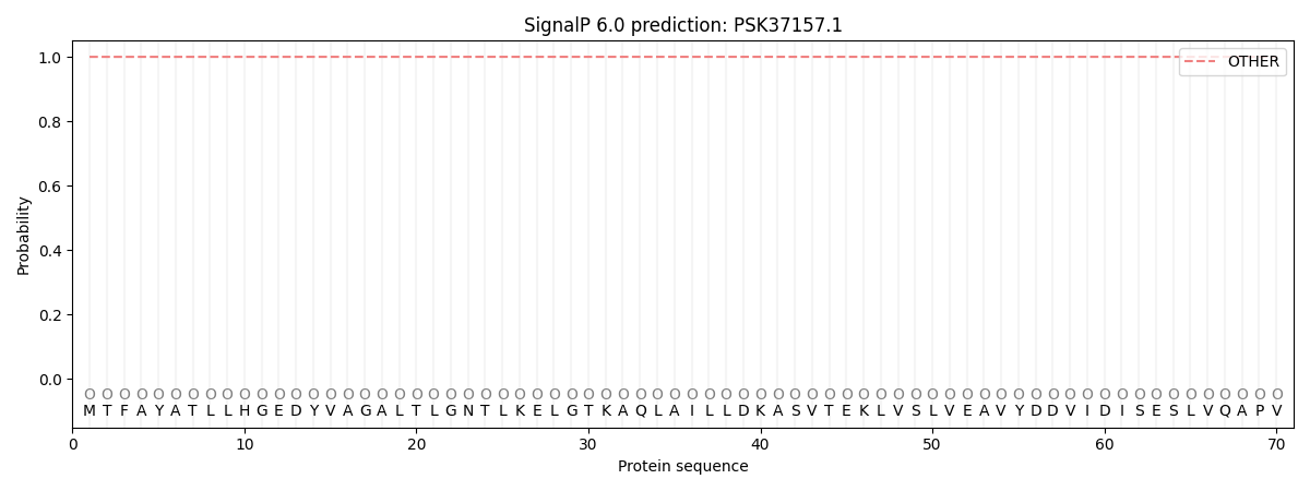
This protein is predicted as OTHER

| Other | SP_Sec_SPI | CS Position |
|---|---|---|
| 1.000044 | 0.000001 |
