You are browsing environment: FUNGIDB
CAZyme Information: PPTG_11791-t26_5-p1
You are here: Home > Sequence: PPTG_11791-t26_5-p1
Basic Information |
Genomic context |
Full Sequence |
Enzyme annotations |
CAZy signature domains |
CDD domains |
CAZyme hits |
PDB hits |
Swiss-Prot hits |
SignalP and Lipop annotations |
TMHMM annotations
Basic Information help
| Species | Phytophthora parasitica | |||||||||||
|---|---|---|---|---|---|---|---|---|---|---|---|---|
| Lineage | Oomycota; NA; ; Peronosporaceae; Phytophthora; Phytophthora parasitica | |||||||||||
| CAZyme ID | PPTG_11791-t26_5-p1 | |||||||||||
| CAZy Family | GH5 | |||||||||||
| CAZyme Description | hypothetical protein, variant 1 | |||||||||||
| CAZyme Property |
|
|||||||||||
| Genome Property |
|
|||||||||||
| Gene Location | ||||||||||||
CAZyme Signature Domains help
| Family | Start | End | Evalue | family coverage |
|---|---|---|---|---|
| CBM47 | 1013 | 1152 | 1.2e-18 | 0.9609375 |
CDD Domains download full data without filtering help
| Cdd ID | Domain | E-Value | qStart | qEnd | sStart | sEnd | Domain Description |
|---|---|---|---|---|---|---|---|
| 227511 | ATS1 | 9.04e-28 | 705 | 981 | 94 | 374 | Alpha-tubulin suppressor and related RCC1 domain-containing proteins [Cell cycle control, cell division, chromosome partitioning, Cytoskeleton]. |
| 227511 | ATS1 | 7.46e-19 | 702 | 986 | 34 | 323 | Alpha-tubulin suppressor and related RCC1 domain-containing proteins [Cell cycle control, cell division, chromosome partitioning, Cytoskeleton]. |
| 238008 | EFh | 3.04e-16 | 60 | 119 | 3 | 62 | EF-hand, calcium binding motif; A diverse superfamily of calcium sensors and calcium signal modulators; most examples in this alignment model have 2 active canonical EF hands. Ca2+ binding induces a conformational change in the EF-hand motif, leading to the activation or inactivation of target proteins. EF-hands tend to occur in pairs or higher copy numbers. |
| 320055 | EFh_PEF_Group_I | 2.48e-15 | 13 | 117 | 1 | 91 | Penta-EF hand, calcium binding motifs, found in Group I PEF proteins. The family corresponds to Group I PEF proteins that have been found not only in higher animals but also in lower animals, plants, fungi and protists. Group I PEF proteins include apoptosis-linked gene 2 protein (ALG-2), peflin and similar proteins. ALG-2, also termed programmed cell death protein 6 (PDCD6), is a widely expressed calcium-binding modulator protein associated with cell proliferation and death, as well as cell survival. It forms a homodimer in the cell or a heterodimer with its closest paralog peflin. Among the PEF proteins, ALG-2 can bind three Ca2+ ions through its EF1, EF3, and EF5 hands, where it is unique in that its EF5 hand binds Ca2+ ion in a canonical coordination. Peflin is a ubiquitously expressed 30-kD PEF protein containing five EF-hand motifs in its C-terminal domain and a longer N-terminal hydrophobic domain (NHB domain) than any other member of the PEF family. The NHB domain harbors nine repeats of a nonapeptide (A/PPGGPYGGP). Peflin may modulate the function of ALG-2 in Ca2+ signaling. It exists only as a heterodimer with ALG-2, and binds two Ca2+ ions through its EF1 and EF3 hands. Its additional EF5 hand is unpaired and does not bind Ca2+ ion but mediates the heterodimerization with ALG-2. The dissociation of heterodimer occurs in the presence of Ca2+. |
| 227511 | ATS1 | 1.31e-13 | 794 | 991 | 68 | 275 | Alpha-tubulin suppressor and related RCC1 domain-containing proteins [Cell cycle control, cell division, chromosome partitioning, Cytoskeleton]. |
CAZyme Hits help
| Hit ID | E-Value | Query Start | Query End | Hit Start | Hit End |
|---|---|---|---|---|---|
| 2.86e-15 | 719 | 977 | 91 | 326 | |
| 1.00e-13 | 719 | 977 | 93 | 328 | |
| 4.33e-13 | 1003 | 1157 | 203 | 340 | |
| 4.33e-13 | 1003 | 1157 | 203 | 340 | |
| 6.82e-13 | 719 | 977 | 106 | 339 |
PDB Hits download full data without filtering help
| Hit ID | E-Value | Query Start | Query End | Hit Start | Hit End | Description |
|---|---|---|---|---|---|---|
| 1.15e-28 | 709 | 1000 | 105 | 372 | Chain A, Ultraviolet-B receptor UVR8 [Arabidopsis thaliana],6XZM_B Chain B, Ultraviolet-B receptor UVR8 [Arabidopsis thaliana] |
|
| 1.15e-28 | 709 | 1000 | 105 | 372 | Chain A, Ultraviolet-B receptor UVR8 [Arabidopsis thaliana],6XZN_B Chain B, Ultraviolet-B receptor UVR8 [Arabidopsis thaliana] |
|
| 2.69e-28 | 709 | 1000 | 102 | 369 | Crystal structure of the W285F mutant of UVB-resistance protein UVR8 [Arabidopsis thaliana],4DNV_B Crystal structure of the W285F mutant of UVB-resistance protein UVR8 [Arabidopsis thaliana],4DNV_C Crystal structure of the W285F mutant of UVB-resistance protein UVR8 [Arabidopsis thaliana],4DNV_D Crystal structure of the W285F mutant of UVB-resistance protein UVR8 [Arabidopsis thaliana] |
|
| 2.88e-28 | 709 | 1000 | 102 | 369 | Crystal structure of UVB-resistance protein UVR8 [Arabidopsis thaliana],4DNW_B Crystal structure of UVB-resistance protein UVR8 [Arabidopsis thaliana] |
|
| 3.04e-28 | 709 | 1000 | 101 | 368 | Crystal structure of UVB photoreceptor UVR8 from Arabidopsis thaliana and UV-induced structural changes at 120K [Arabidopsis thaliana],4NAA_B Crystal structure of UVB photoreceptor UVR8 from Arabidopsis thaliana and UV-induced structural changes at 120K [Arabidopsis thaliana],4NAA_C Crystal structure of UVB photoreceptor UVR8 from Arabidopsis thaliana and UV-induced structural changes at 120K [Arabidopsis thaliana],4NAA_D Crystal structure of UVB photoreceptor UVR8 from Arabidopsis thaliana and UV-induced structural changes at 120K [Arabidopsis thaliana],4NBM_A Crystal structure of UVB photoreceptor UVR8 and light-induced structural changes at 180K [Arabidopsis thaliana],4NBM_B Crystal structure of UVB photoreceptor UVR8 and light-induced structural changes at 180K [Arabidopsis thaliana],4NBM_C Crystal structure of UVB photoreceptor UVR8 and light-induced structural changes at 180K [Arabidopsis thaliana],4NBM_D Crystal structure of UVB photoreceptor UVR8 and light-induced structural changes at 180K [Arabidopsis thaliana],6DD7_A Crystal structure of plant UVB photoreceptor UVR8 from in situ serial Laue diffraction [Arabidopsis thaliana],6DD7_B Crystal structure of plant UVB photoreceptor UVR8 from in situ serial Laue diffraction [Arabidopsis thaliana],6DD7_C Crystal structure of plant UVB photoreceptor UVR8 from in situ serial Laue diffraction [Arabidopsis thaliana],6DD7_D Crystal structure of plant UVB photoreceptor UVR8 from in situ serial Laue diffraction [Arabidopsis thaliana] |
Swiss-Prot Hits download full data without filtering help
| Hit ID | E-Value | Query Start | Query End | Hit Start | Hit End | Description |
|---|---|---|---|---|---|---|
| 3.81e-28 | 710 | 977 | 15 | 265 | E3 ISG15--protein ligase Herc6 OS=Mus musculus OX=10090 GN=Herc6 PE=2 SV=1 |
|
| 4.24e-27 | 709 | 1000 | 113 | 380 | Ultraviolet-B receptor UVR8 OS=Arabidopsis thaliana OX=3702 GN=UVR8 PE=1 SV=1 |
|
| 9.20e-26 | 719 | 984 | 25 | 273 | Probable E3 ubiquitin-protein ligase HERC6 OS=Homo sapiens OX=9606 GN=HERC6 PE=1 SV=2 |
|
| 6.51e-25 | 693 | 992 | 2992 | 3275 | Probable E3 ubiquitin-protein ligase HERC2 OS=Drosophila melanogaster OX=7227 GN=HERC2 PE=1 SV=3 |
|
| 1.11e-24 | 693 | 1073 | 2966 | 3351 | E3 ubiquitin-protein ligase HERC2 OS=Mus musculus OX=10090 GN=Herc2 PE=1 SV=3 |
SignalP and Lipop Annotations help
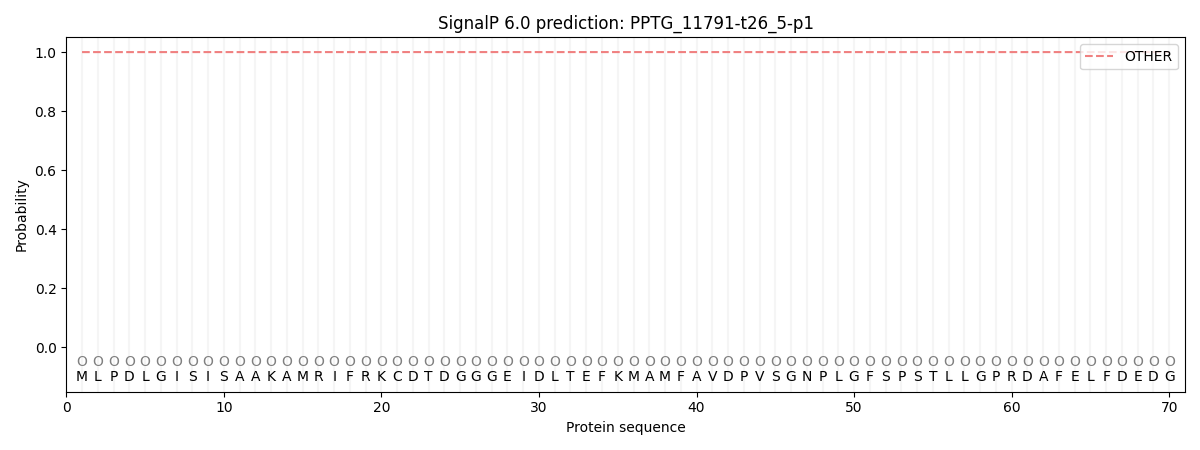
This protein is predicted as OTHER

| Other | SP_Sec_SPI | CS Position |
|---|---|---|
| 1.000051 | 0.000000 |
