You are browsing environment: FUNGIDB
CAZyme Information: PNEJI1_001875-t26_1-p1
You are here: Home > Sequence: PNEJI1_001875-t26_1-p1
Basic Information |
Genomic context |
Full Sequence |
Enzyme annotations |
CAZy signature domains |
CDD domains |
CAZyme hits |
PDB hits |
Swiss-Prot hits |
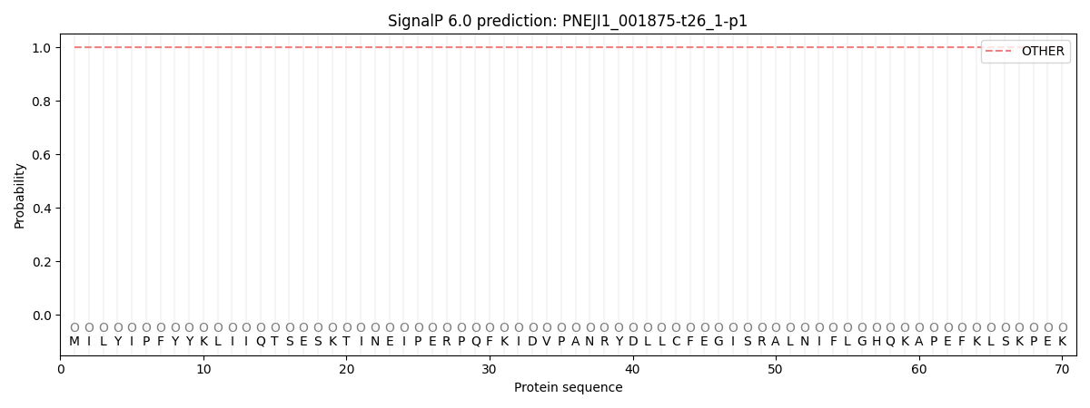
SignalP and Lipop annotations |
TMHMM annotations
Basic Information help
| Species | Pneumocystis jirovecii | |||||||||||
|---|---|---|---|---|---|---|---|---|---|---|---|---|
| Lineage | Ascomycota; Pneumocystidomycetes; ; Pneumocystidaceae; Pneumocystis; Pneumocystis jirovecii | |||||||||||
| CAZyme ID | PNEJI1_001875-t26_1-p1 | |||||||||||
| CAZy Family | GT22 | |||||||||||
| CAZyme Description | unspecified product | |||||||||||
| CAZyme Property |
|
|||||||||||
| Genome Property |
|
|||||||||||
| Gene Location | ||||||||||||
CAZyme Signature Domains help
| Family | Start | End | Evalue | family coverage |
|---|---|---|---|---|
| GT8 | 451 | 606 | 3.3e-22 | 0.7431906614785992 |
CDD Domains download full data without filtering help
| Cdd ID | Domain | E-Value | qStart | qEnd | sStart | sEnd | Domain Description |
|---|---|---|---|---|---|---|---|
| 215149 | PLN02265 | 1.34e-163 | 30 | 443 | 67 | 564 | probable phenylalanyl-tRNA synthetase beta chain |
| 273096 | pheT_arch | 2.11e-98 | 26 | 445 | 40 | 520 | phenylalanyl-tRNA synthetase, beta subunit. Every known example of the phenylalanyl-tRNA synthetase, except the monomeric form of mitochondrial, is an alpha 2 beta 2 heterotetramer. The beta subunits break into two subfamilies that are considerably different in sequence, length, and pattern of gaps. This model represents the subfamily that includes the beta subunit from eukaryotic cytosol, the Archaea, and spirochetes. [Protein synthesis, tRNA aminoacylation] |
| 236592 | pheT | 6.16e-91 | 23 | 439 | 36 | 512 | phenylalanine--tRNA ligase subunit beta. |
| 223150 | PheT | 3.00e-76 | 94 | 441 | 178 | 512 | Phenylalanyl-tRNA synthetase beta subunit [Translation, ribosomal structure and biogenesis]. |
| 133018 | GT8_Glycogenin | 2.72e-46 | 450 | 611 | 14 | 188 | Glycogenin belongs the GT 8 family and initiates the biosynthesis of glycogen. Glycogenin initiates the biosynthesis of glycogen by incorporating glucose residues through a self-glucosylation reaction at a Tyr residue, and then acts as substrate for chain elongation by glycogen synthase and branching enzyme. It contains a conserved DxD motif and an N-terminal beta-alpha-beta Rossmann-like fold that are common to the nucleotide-binding domains of most glycosyltransferases. The DxD motif is essential for coordination of the catalytic divalent cation, most commonly Mn2+. Glycogenin can be classified as a retaining glycosyltransferase, based on the relative anomeric stereochemistry of the substrate and product in the reaction catalyzed. It is placed in glycosyltransferase family 8 which includes lipopolysaccharide glucose and galactose transferases and galactinol synthases. |
CAZyme Hits help
| Hit ID | E-Value | Query Start | Query End | Hit Start | Hit End |
|---|---|---|---|---|---|
| 0.0 | 15 | 612 | 39 | 770 | |
| 6.79e-36 | 451 | 612 | 25 | 200 | |
| 1.26e-35 | 451 | 612 | 16 | 190 | |
| 2.15e-33 | 451 | 612 | 21 | 195 | |
| 2.20e-33 | 451 | 612 | 21 | 197 |
PDB Hits download full data without filtering help
| Hit ID | E-Value | Query Start | Query End | Hit Start | Hit End | Description |
|---|---|---|---|---|---|---|
| 2.12e-105 | 30 | 450 | 63 | 563 | Crystal structure of Homo Sapiens cytoplasmic Phenylalanyl-tRNA synthetase [Homo sapiens],3L4G_D Crystal structure of Homo Sapiens cytoplasmic Phenylalanyl-tRNA synthetase [Homo sapiens],3L4G_F Crystal structure of Homo Sapiens cytoplasmic Phenylalanyl-tRNA synthetase [Homo sapiens],3L4G_H Crystal structure of Homo Sapiens cytoplasmic Phenylalanyl-tRNA synthetase [Homo sapiens],3L4G_J Crystal structure of Homo Sapiens cytoplasmic Phenylalanyl-tRNA synthetase [Homo sapiens],3L4G_L Crystal structure of Homo Sapiens cytoplasmic Phenylalanyl-tRNA synthetase [Homo sapiens],3L4G_N Crystal structure of Homo Sapiens cytoplasmic Phenylalanyl-tRNA synthetase [Homo sapiens],3L4G_P Crystal structure of Homo Sapiens cytoplasmic Phenylalanyl-tRNA synthetase [Homo sapiens] |
|
| 7.33e-52 | 30 | 445 | 47 | 585 | Chain B, Phenylalanyl-tRNA synthetase beta subunit [Plasmodium falciparum],7DPI_D Chain D, Phenylalanyl-tRNA synthetase beta subunit [Plasmodium falciparum] |
|
| 2.13e-46 | 26 | 445 | 44 | 580 | Chain B, Phenylalanyl-tRNA synthetase beta chain, putative [Plasmodium vivax] |
|
| 9.84e-30 | 422 | 611 | 11 | 219 | Crystal Structure of Human Glycogenin-1 (GYG1) complexed with manganese [Homo sapiens] |
|
| 1.14e-29 | 451 | 611 | 19 | 198 | Crystal Structure of Human Glycogenin-1 (GYG1), apo form [Homo sapiens],3QVB_A Crystal Structure of Human Glycogenin-1 (GYG1) complexed with manganese and UDP [Homo sapiens],3U2W_A Crystal Structure of Human Glycogenin-1 (GYG1) complexed with manganese and glucose or a glucal species [Homo sapiens],3U2W_B Crystal Structure of Human Glycogenin-1 (GYG1) complexed with manganese and glucose or a glucal species [Homo sapiens] |
Swiss-Prot Hits download full data without filtering help
| Hit ID | E-Value | Query Start | Query End | Hit Start | Hit End | Description |
|---|---|---|---|---|---|---|
| 2.67e-139 | 26 | 441 | 49 | 548 | Phenylalanine--tRNA ligase beta subunit OS=Schizosaccharomyces pombe (strain 972 / ATCC 24843) OX=284812 GN=frs1 PE=3 SV=1 |
|
| 1.65e-138 | 7 | 441 | 29 | 551 | Phenylalanine--tRNA ligase beta subunit OS=Candida albicans OX=5476 GN=FRS1 PE=2 SV=3 |
|
| 1.30e-122 | 26 | 447 | 48 | 560 | Phenylalanine--tRNA ligase beta subunit OS=Saccharomyces cerevisiae (strain ATCC 204508 / S288c) OX=559292 GN=FRS1 PE=1 SV=3 |
|
| 1.09e-104 | 30 | 450 | 63 | 563 | Phenylalanine--tRNA ligase beta subunit OS=Homo sapiens OX=9606 GN=FARSB PE=1 SV=3 |
|
| 3.31e-103 | 30 | 450 | 63 | 563 | Phenylalanine--tRNA ligase beta subunit OS=Pongo abelii OX=9601 GN=FARSB PE=2 SV=1 |
SignalP and Lipop Annotations help
This protein is predicted as OTHER

| Other | SP_Sec_SPI | CS Position |
|---|---|---|
| 1.000041 | 0.000001 |
