You are browsing environment: FUNGIDB
CAZyme Information: PMAA_023420-t26_1-p1
You are here: Home > Sequence: PMAA_023420-t26_1-p1
Basic Information |
Genomic context |
Full Sequence |
Enzyme annotations |
CAZy signature domains |
CDD domains |
CAZyme hits |
PDB hits |
Swiss-Prot hits |
SignalP and Lipop annotations |
TMHMM annotations
Basic Information help
| Species | Talaromyces marneffei | |||||||||||
|---|---|---|---|---|---|---|---|---|---|---|---|---|
| Lineage | Ascomycota; Eurotiomycetes; ; Trichocomaceae; Talaromyces; Talaromyces marneffei | |||||||||||
| CAZyme ID | PMAA_023420-t26_1-p1 | |||||||||||
| CAZy Family | CE4 | |||||||||||
| CAZyme Description | conserved hypothetical protein | |||||||||||
| CAZyme Property |
|
|||||||||||
| Genome Property |
|
|||||||||||
| Gene Location | ||||||||||||
CAZyme Signature Domains help
| Family | Start | End | Evalue | family coverage |
|---|---|---|---|---|
| AA7 | 42 | 452 | 5.8e-51 | 0.9606986899563319 |
CDD Domains download full data without filtering help
| Cdd ID | Domain | E-Value | qStart | qEnd | sStart | sEnd | Domain Description |
|---|---|---|---|---|---|---|---|
| 223354 | GlcD | 2.36e-30 | 29 | 225 | 21 | 226 | FAD/FMN-containing dehydrogenase [Energy production and conversion]. |
| 396238 | FAD_binding_4 | 4.99e-30 | 44 | 175 | 1 | 135 | FAD binding domain. This family consists of various enzymes that use FAD as a co-factor, most of the enzymes are similar to oxygen oxidoreductase. One of the enzymes Vanillyl-alcohol oxidase (VAO) has a solved structure, the alignment includes the FAD binding site, called the PP-loop, between residues 99-110. The FAD molecule is covalently bound in the known structure, however the residue that links to the FAD is not in the alignment. VAO catalyzes the oxidation of a wide variety of substrates, ranging form aromatic amines to 4-alkylphenols. Other members of this family include D-lactate dehydrogenase, this enzyme catalyzes the conversion of D-lactate to pyruvate using FAD as a co-factor; mitomycin radical oxidase, this enzyme oxidizes the reduced form of mitomycins and is involved in mitomycin resistance. This family includes MurB an UDP-N-acetylenolpyruvoylglucosamine reductase enzyme EC:1.1.1.158. This enzyme is involved in the biosynthesis of peptidoglycan. |
| 410683 | CYP57A1-like | 1.56e-23 | 430 | 509 | 340 | 425 | cytochrome P450 family 57, subfamily A, polypeptide 1 and similar cytochrome P450s. This family is composed of fungal cytochrome P450s including: Nectria haematococca cytochrome P450 57A1 (CYP57A1), also called pisatin demethylase, which detoxifies the phytoalexin pisatin; Penicillium aethiopicum P450 monooxygenase gsfF, also called griseofulvin synthesis protein F, which catalyzes the coupling of orcinol and phloroglucinol rings in griseophenone B to form desmethyl-dehydrogriseofulvin A during the biosynthesis of griseofulvin, a spirocyclic fungal natural product used to treat dermatophyte infections; and Penicillium aethiopicum P450 monooxygenase vrtE, also called viridicatumtoxin synthesis protein E, which catalyzes hydroxylation at C5 of the polyketide backbone during the biosynthesis of viridicatumtoxin, a tetracycline-like fungal meroterpenoid. The CYP57A1-like family belongs to the large cytochrome P450 (P450, CYP) superfamily of heme-containing proteins that catalyze a variety of oxidative reactions of a large number of structurally different endogenous and exogenous compounds in organisms from all major domains of life. CYPs bind their diverse ligands in a buried, hydrophobic active site, which is accessed through a substrate access channel formed by two flexible helices and their connecting loop. |
| 273751 | FAD_lactone_ox | 4.26e-14 | 42 | 205 | 13 | 180 | sugar 1,4-lactone oxidases. This model represents a family of at least two different sugar 1,4 lactone oxidases, both involved in synthesizing ascorbic acid or a derivative. These include L-gulonolactone oxidase (EC 1.1.3.8) from rat and D-arabinono-1,4-lactone oxidase (EC 1.1.3.37) from Saccharomyces cerevisiae. Members are proposed to have the cofactor FAD covalently bound at a site specified by Prosite motif PS00862; OX2_COVAL_FAD; 1. |
| 223882 | MurB | 1.28e-07 | 47 | 205 | 24 | 182 | UDP-N-acetylenolpyruvoylglucosamine reductase [Cell wall/membrane/envelope biogenesis]. |
CAZyme Hits help
| Hit ID | E-Value | Query Start | Query End | Hit Start | Hit End |
|---|---|---|---|---|---|
| 4.38e-21 | 22 | 451 | 43 | 483 | |
| 4.38e-21 | 22 | 451 | 43 | 483 | |
| 5.87e-21 | 22 | 451 | 43 | 483 | |
| 7.80e-20 | 44 | 483 | 90 | 544 | |
| 8.15e-20 | 24 | 451 | 46 | 484 |
PDB Hits download full data without filtering help
| Hit ID | E-Value | Query Start | Query End | Hit Start | Hit End | Description |
|---|---|---|---|---|---|---|
| 2.28e-21 | 44 | 224 | 39 | 224 | Crystal structure of 6-hydoxy-D-nicotine oxidase from Arthrobacter nicotinovorans. Crystal Form 3 (P1) [Paenarthrobacter nicotinovorans],2BVF_B Crystal structure of 6-hydoxy-D-nicotine oxidase from Arthrobacter nicotinovorans. Crystal Form 3 (P1) [Paenarthrobacter nicotinovorans],2BVG_A Crystal structure of 6-hydoxy-D-nicotine oxidase from Arthrobacter nicotinovorans. Crystal Form 1 (P21) [Paenarthrobacter nicotinovorans],2BVG_B Crystal structure of 6-hydoxy-D-nicotine oxidase from Arthrobacter nicotinovorans. Crystal Form 1 (P21) [Paenarthrobacter nicotinovorans],2BVG_C Crystal structure of 6-hydoxy-D-nicotine oxidase from Arthrobacter nicotinovorans. Crystal Form 1 (P21) [Paenarthrobacter nicotinovorans],2BVG_D Crystal structure of 6-hydoxy-D-nicotine oxidase from Arthrobacter nicotinovorans. Crystal Form 1 (P21) [Paenarthrobacter nicotinovorans],2BVH_A Crystal structure of 6-hydoxy-D-nicotine oxidase from Arthrobacter nicotinovorans. Crystal Form 2 (P21) [Paenarthrobacter nicotinovorans],2BVH_B Crystal structure of 6-hydoxy-D-nicotine oxidase from Arthrobacter nicotinovorans. Crystal Form 2 (P21) [Paenarthrobacter nicotinovorans],2BVH_C Crystal structure of 6-hydoxy-D-nicotine oxidase from Arthrobacter nicotinovorans. Crystal Form 2 (P21) [Paenarthrobacter nicotinovorans],2BVH_D Crystal structure of 6-hydoxy-D-nicotine oxidase from Arthrobacter nicotinovorans. Crystal Form 2 (P21) [Paenarthrobacter nicotinovorans] |
|
| 3.48e-21 | 20 | 452 | 24 | 463 | Physcomitrella patens BBE-like 1 wild-type [Physcomitrium patens],6EO4_B Physcomitrella patens BBE-like 1 wild-type [Physcomitrium patens] |
|
| 3.48e-21 | 20 | 452 | 24 | 463 | Physcomitrella patens BBE-like 1 variant D396N [Physcomitrium patens],6EO5_B Physcomitrella patens BBE-like 1 variant D396N [Physcomitrium patens] |
|
| 7.32e-17 | 24 | 228 | 28 | 249 | Chain A, MaDA [Morus alba],6JQH_B Chain B, MaDA [Morus alba] |
|
| 1.31e-15 | 36 | 203 | 70 | 246 | Structure of BBE-like #28 from Arabidopsis thaliana [Arabidopsis thaliana],5D79_B Structure of BBE-like #28 from Arabidopsis thaliana [Arabidopsis thaliana] |
Swiss-Prot Hits download full data without filtering help
| Hit ID | E-Value | Query Start | Query End | Hit Start | Hit End | Description |
|---|---|---|---|---|---|---|
| 3.67e-44 | 17 | 458 | 69 | 501 | FAD-linked oxidoreductase aurO OS=Gibberella zeae (strain ATCC MYA-4620 / CBS 123657 / FGSC 9075 / NRRL 31084 / PH-1) OX=229533 GN=aurO PE=1 SV=1 |
|
| 8.37e-25 | 44 | 451 | 48 | 441 | FAD-linked oxidoreductase DDB_G0289697 OS=Dictyostelium discoideum OX=44689 GN=DDB_G0289697 PE=2 SV=1 |
|
| 1.16e-20 | 44 | 224 | 38 | 223 | (R)-6-hydroxynicotine oxidase OS=Paenarthrobacter nicotinovorans OX=29320 GN=6-hdno PE=1 SV=2 |
|
| 4.83e-20 | 24 | 218 | 18 | 217 | FAD-linked oxidoreductase pyvE OS=Aspergillus violaceofuscus (strain CBS 115571) OX=1450538 GN=pyvE PE=3 SV=1 |
|
| 1.05e-18 | 50 | 451 | 65 | 472 | FAD-linked oxidoreductase afoF OS=Emericella nidulans (strain FGSC A4 / ATCC 38163 / CBS 112.46 / NRRL 194 / M139) OX=227321 GN=afoF PE=1 SV=1 |
SignalP and Lipop Annotations help
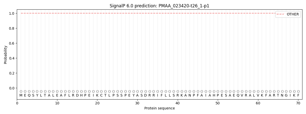
This protein is predicted as OTHER

| Other | SP_Sec_SPI | CS Position |
|---|---|---|
| 1.000058 | 0.000000 |
