You are browsing environment: FUNGIDB
CAZyme Information: PIW_T001129-RA-p1
You are here: Home > Sequence: PIW_T001129-RA-p1
Basic Information |
Genomic context |
Full Sequence |
Enzyme annotations |
CAZy signature domains |
CDD domains |
CAZyme hits |
PDB hits |
Swiss-Prot hits |
SignalP and Lipop annotations |
TMHMM annotations
Basic Information help
| Species | Globisporangium iwayamae | |||||||||||
|---|---|---|---|---|---|---|---|---|---|---|---|---|
| Lineage | Oomycota; NA; ; Pythiaceae; Globisporangium; Globisporangium iwayamae | |||||||||||
| CAZyme ID | PIW_T001129-RA-p1 | |||||||||||
| CAZy Family | AA17 | |||||||||||
| CAZyme Description | Alcohol dehydrogenase | |||||||||||
| CAZyme Property |
|
|||||||||||
| Genome Property |
|
|||||||||||
| Gene Location | Start: 10292; End:14867 Strand: - | |||||||||||
Full Sequence Download help
| MCARKAGQRS RCARAFSLLR WCGTSWRQCM HSLRVARSLL SVLSPQLQAL VEASLGDSLA | 60 |
| RSSTTTLSQS SAGASAALHT ATAAESDNPS PTAEKRAATT TTLSTELRDG AQHDRVPVSF | 120 |
| KLNPSELFVL RHPASPGFVV KQNFLGSTGA TAVRDALLAL AETETFQEAK VGHGDHLRSA | 180 |
| RAVRGDRIHW LKRPGDLSQS AGSDGPHPAI LHLMESVESL VYGVKSAVPS LNIRNVTSTQ | 240 |
| LAIFPGGGAR FVRHADAYSS AHHEDASRSS PSSLDGLVRV LTCVYYLNDH WEPNHGGQLR | 300 |
| VYVDGSSSSS SSSSSSLVAA AAGVAHWDVA PALDTLVVFR GLDVEHEVLP AFRERMALTI | 360 |
| WYYGRVESQP ATHPSRIRIG ICLQDDNDDD TRQYLEDKYS ADQIDSHMRF RAGWDAFLIN | 420 |
| QLEKCPSAKP ILTTYPLGYT LPNNVSTDIR PTLLCASSFD EHGILRQASK TLTRVSPVPL | 480 |
| ASSFWAAGFA FSSARVISEV PYDESLRFLF FGEEASMNAR LWTAGWDFFT PGESVVYHLW | 540 |
| TRSYRRVFQE IEDQETVKWR AASQQYVKTL LTHSSNADSE ASGSEQPELS AGKYSLGVER | 600 |
| SLSTYQSRIG VSFEKQKIRW EAEWGNLDPI HFELSARAAD MTETPPAVIP PTFRAYQYEQ | 660 |
| CGDPFEEIKL RTGIRQTSLQ PHDVRIQVIS AALNPVDYKL VEGSRHAQLG RKPSHETPFT | 720 |
| VGFDLAGVVV EVGLSVADLK VGDHVFAMMP WLKFGSFAEY AVVNEEYVAH KPEALSFDEA | 780 |
| AGVPLAATTS FQVLFQHAKL QPGERVLILG GSSATGIFGI QLAKAAGAYV IATTSFRNTT | 840 |
| FVEGLDADQV IDYTQESWGD VLAQHSVDVI YDCGMEPDAW SRVAQQVLKK HTGRFVSLLP | 900 |
| SPSNNSRQIE SRSGATNLGQ IMVNPTAAHL REIAKLFDNK SLVAPVDSVY EFDELFDAIR | 960 |
| KLKTGRARGK LILHVHSPPP HHYPADH | 987 |
CAZyme Signature Domains help
| Family | Start | End | Evalue | family coverage |
|---|---|---|---|---|
| GT60 | 403 | 614 | 1.6e-62 | 0.6515151515151515 |
CDD Domains download full data without filtering help
| Cdd ID | Domain | E-Value | qStart | qEnd | sStart | sEnd | Domain Description |
|---|---|---|---|---|---|---|---|
| 176191 | MDR_like_2 | 7.25e-99 | 654 | 973 | 2 | 309 | alcohol dehydrogenase and quinone reductase-like medium chain degydrogenases/reductases. Members identified as zinc-dependent alcohol dehydrogenases and quinone oxidoreductase. QOR catalyzes the conversion of a quinone + NAD(P)H to a hydroquinone + NAD(P)+. Quinones are cyclic diones derived from aromatic compounds. Membrane bound QOR actin the respiratory chains of bacteria and mitochondria, while soluble QOR acts to protect from toxic quinones (e.g. DT-diaphorase) or as a soluble eye-lens protein in some vertebrates (e.g. zeta-crystalin). QOR reduces quinones through a semi-quinone intermediate via a NAD(P)H-dependent single electron transfer. QOR is a member of the medium chain dehydrogenase/reductase family, but lacks the zinc-binding sites of the prototypical alcohol dehydrogenases of this group. NAD(P)(H)-dependent oxidoreductases are the major enzymes in the interconversion of alcohols and aldehydes, or ketones. Alcohol dehydrogenase in the liver converts ethanol and NAD+ to acetaldehyde and NADH, while in yeast and some other microorganisms ADH catalyzes the conversion acetaldehyde to ethanol in alcoholic fermentation. ADH is a member of the medium chain alcohol dehydrogenase family (MDR), which has a NAD(P)(H)-binding domain in a Rossmann fold of a beta-alpha form. The NAD(H)-binding region is comprised of 2 structurally similar halves, each of which contacts a mononucleotide. A GxGxxG motif after the first mononucleotide contact half allows the close contact of the coenzyme with the ADH backbone. The N-terminal catalytic domain has a distant homology to GroES. These proteins typically form dimers (typically higher plants, mammals) or tetramers (yeast, bacteria), and have 2 tightly bound zinc atoms per subunit, a catalytic zinc at the active site and a structural zinc in a lobe of the catalytic domain. NAD(H) binding occurs in the cleft between the catalytic and coenzyme-binding domains at the active site, and coenzyme binding induces a conformational closing of this cleft. Coenzyme binding typically precedes and contributes to substrate binding. In human ADH catalysis, the zinc ion helps coordinate the alcohol, followed by deprotonation of a histidine, the ribose of NAD, a serine, then the alcohol, which allows the transfer of a hydride to NAD+, creating NADH and a zinc-bound aldehyde or ketone. In yeast and some bacteria, the active site zinc binds an aldehyde, polarizing it, and leading to the reverse reaction. |
| 176228 | MDR1 | 3.38e-96 | 681 | 973 | 26 | 319 | Medium chain dehydrogenases/reductase (MDR)/zinc-dependent alcohol dehydrogenase-like family. This group is a member of the medium chain dehydrogenases/reductase (MDR)/zinc-dependent alcohol dehydrogenase-like family, but lacks the zinc-binding sites of the zinc-dependent alcohol dehydrogenases. The medium chain dehydrogenases/reductase (MDR)/zinc-dependent alcohol dehydrogenase-like family, which contains the zinc-dependent alcohol dehydrogenase (ADH-Zn) and related proteins, is a diverse group of proteins related to the first identified member, class I mammalian ADH. MDRs display a broad range of activities and are distinguished from the smaller short chain dehydrogenases (~ 250 amino acids vs. the ~ 350 amino acids of the MDR). The MDR proteins have 2 domains: a C-terminal NAD(P)-binding Rossmann fold domain of a beta-alpha form and an N-terminal catalytic domain with distant homology to GroES. The MDR group contains a host of activities, including the founding alcohol dehydrogenase (ADH), quinone reductase, sorbitol dehydrogenase, formaldehyde dehydrogenase, butanediol DH, ketose reductase, cinnamyl reductase, and numerous others. The zinc-dependent alcohol dehydrogenases (ADHs) catalyze the NAD(P)(H)-dependent interconversion of alcohols to aldehydes or ketones. Active site zinc has a catalytic role, while structural zinc aids in stability. ADH-like proteins typically form dimers (typically higher plants, mammals) or tetramers (yeast, bacteria), and generally have 2 tightly bound zinc atoms per subunit. The active site zinc is coordinated by a histidine, two cysteines, and a water molecule. The second zinc seems to play a structural role, affects subunit interactions, and is typically coordinated by 4 cysteines. |
| 176210 | RTN4I1 | 9.14e-66 | 654 | 972 | 2 | 348 | Human Reticulon 4 Interacting Protein 1. Human Reticulon 4 Interacting Protein 1 is a member of the medium chain dehydrogenase/ reductase (MDR) family. Riticulons are endoplasmic reticulum associated proteins involved in membrane trafficking and neuroendocrine secretion. The MDR/zinc-dependent alcohol dehydrogenase-like family, which contains the zinc-dependent alcohol dehydrogenase (ADH-Zn) and related proteins, is a diverse group of proteins related to the first identified member, class I mammalian ADH. MDRs display a broad range of activities and are distinguished from the smaller short chain dehydrogenases (~ 250 amino acids vs. the ~ 350 amino acids of the MDR). The MDR proteins have 2 domains: a C-terminal NAD(P) binding-Rossmann fold domain of a beta-alpha form and an N-terminal catalytic domain with distant homology to GroES. |
| 176211 | enoyl_reductase_like | 9.97e-66 | 678 | 975 | 23 | 339 | enoyl_reductase_like. Member identified as possible enoyl reductase of the MDR family. 2-enoyl thioester reductase (ETR) catalyzes the NADPH-dependent dependent conversion of trans-2-enoyl acyl carrier protein/coenzyme A (ACP/CoA) to acyl-(ACP/CoA) in fatty acid synthesis. 2-enoyl thioester reductase activity has been linked in Candida tropicalis as essential in maintaining mitiochondrial respiratory function. This ETR family is a part of the medium chain dehydrogenase/reductase family, but lack the zinc coordination sites characteristic of the alcohol dehydrogenases in this family. NAD(P)(H)-dependent oxidoreductases are the major enzymes in the interconversion of alcohols and aldehydes, or ketones. Alcohol dehydrogenase in the liver converts ethanol and NAD+ to acetaldehyde and NADH, while in yeast and some other microorganisms ADH catalyzes the conversion acetaldehyde to ethanol in alcoholic fermentation. ADH is a member of the medium chain alcohol dehydrogenase family (MDR), which has a NAD(P)(H)-binding domain in a Rossmann fold of a beta-alpha form. The NAD(H)-binding region is comprised of 2 structurally similar halves, each of which contacts a mononucleotide. The N-terminal catalytic domain has a distant homology to GroES. These proteins typically form dimers (typically higher plants, mammals) or tetramers (yeast, bacteria), and have 2 tightly bound zinc atoms per subunit, a catalytic zinc at the active site, and a structural zinc in a lobe of the catalytic domain. NAD(H)-binding occurs in the cleft between the catalytic and coenzyme-binding domains at the active site, and coenzyme binding induces a conformational closing of this cleft. Coenzyme binding typically precedes and contributes to substrate binding. Candida tropicalis enoyl thioester reductase (Etr1p) catalyzes the NADPH-dependent reduction of trans-2-enoyl thioesters in mitochondrial fatty acid synthesis. Etr1p forms homodimers with each subunit containing a nucleotide-binding Rossmann fold domain and a catalytic domain. |
| 223677 | Qor | 5.35e-65 | 653 | 975 | 1 | 326 | NADPH:quinone reductase or related Zn-dependent oxidoreductase [Energy production and conversion, General function prediction only]. |
CAZyme Hits help
| Hit ID | E-Value | Query Start | Query End | Hit Start | Hit End |
|---|---|---|---|---|---|
| CCA15104.1|GT60 | 1.14e-110 | 122 | 637 | 89 | 641 |
| UIZ23002.1|GT60 | 8.54e-55 | 479 | 634 | 15 | 163 |
| BDA44136.1|GT60 | 3.34e-52 | 174 | 625 | 97 | 595 |
| CAG4710602.1|GT60 | 1.61e-41 | 127 | 634 | 60 | 661 |
| AAK56291.1|GT60|2.4.1.- | 1.25e-36 | 403 | 558 | 100 | 267 |
PDB Hits download full data without filtering help
| Hit ID | E-Value | Query Start | Query End | Hit Start | Hit End | Description |
|---|---|---|---|---|---|---|
| 4IDA_A | 8.34e-39 | 645 | 974 | 16 | 330 | Structure of the Fragaria x ananassa enone oxidoreductase in its apo form [Fragaria vesca],4IDB_A Structure of the Fragaria x ananassa enone oxidoreductase in complex with NADP+ [Fragaria vesca],4IDC_A Structure of the Fragaria x ananassa enone oxidoreductase in complex with NADPH and HDMF [Fragaria vesca],4IDD_A Structure of the Fragaria x ananassa enone oxidoreductase in complex with NADPH and EHMF [Fragaria vesca],4IDE_A Structure of the Fragaria x ananassa enone oxidoreductase in complex with NADP+ and EDHMF [Fragaria vesca],4IDF_A Structure of the Fragaria x ananassa enone oxidoreductase in complex with NADPH and HMF [Fragaria vesca] |
| 2VN8_A | 2.60e-34 | 681 | 975 | 50 | 374 | Crystal structure of human Reticulon 4 interacting protein 1 in complex with NADPH [Homo sapiens],2VN8_B Crystal structure of human Reticulon 4 interacting protein 1 in complex with NADPH [Homo sapiens] |
| 3TQH_A | 5.48e-31 | 654 | 975 | 8 | 320 | Structure of the quinone oxidoreductase from Coxiella burnetii [Coxiella burnetii] |
| 5A3J_A | 3.32e-29 | 682 | 973 | 34 | 327 | Crystal structure of the chloroplastic gamma-ketol reductase from Arabidopsis thaliana bound to 13-Oxo-9(Z),11(E),15(Z)- octadecatrienoic acid. [Arabidopsis thaliana],5A3J_B Crystal structure of the chloroplastic gamma-ketol reductase from Arabidopsis thaliana bound to 13-Oxo-9(Z),11(E),15(Z)- octadecatrienoic acid. [Arabidopsis thaliana],5A3J_C Crystal structure of the chloroplastic gamma-ketol reductase from Arabidopsis thaliana bound to 13-Oxo-9(Z),11(E),15(Z)- octadecatrienoic acid. [Arabidopsis thaliana],5A3J_D Crystal structure of the chloroplastic gamma-ketol reductase from Arabidopsis thaliana bound to 13-Oxo-9(Z),11(E),15(Z)- octadecatrienoic acid. [Arabidopsis thaliana],5A3J_E Crystal structure of the chloroplastic gamma-ketol reductase from Arabidopsis thaliana bound to 13-Oxo-9(Z),11(E),15(Z)- octadecatrienoic acid. [Arabidopsis thaliana],5A3J_F Crystal structure of the chloroplastic gamma-ketol reductase from Arabidopsis thaliana bound to 13-Oxo-9(Z),11(E),15(Z)- octadecatrienoic acid. [Arabidopsis thaliana],5A3J_G Crystal structure of the chloroplastic gamma-ketol reductase from Arabidopsis thaliana bound to 13-Oxo-9(Z),11(E),15(Z)- octadecatrienoic acid. [Arabidopsis thaliana],5A3J_H Crystal structure of the chloroplastic gamma-ketol reductase from Arabidopsis thaliana bound to 13-Oxo-9(Z),11(E),15(Z)- octadecatrienoic acid. [Arabidopsis thaliana],5A3J_I Crystal structure of the chloroplastic gamma-ketol reductase from Arabidopsis thaliana bound to 13-Oxo-9(Z),11(E),15(Z)- octadecatrienoic acid. [Arabidopsis thaliana],5A3J_J Crystal structure of the chloroplastic gamma-ketol reductase from Arabidopsis thaliana bound to 13-Oxo-9(Z),11(E),15(Z)- octadecatrienoic acid. [Arabidopsis thaliana],5A3J_K Crystal structure of the chloroplastic gamma-ketol reductase from Arabidopsis thaliana bound to 13-Oxo-9(Z),11(E),15(Z)- octadecatrienoic acid. [Arabidopsis thaliana],5A3J_L Crystal structure of the chloroplastic gamma-ketol reductase from Arabidopsis thaliana bound to 13-Oxo-9(Z),11(E),15(Z)- octadecatrienoic acid. [Arabidopsis thaliana],5A3V_A Crystal structure of the chloroplastic gamma-ketol reductase from Arabidopsis thaliana [Arabidopsis thaliana],5A3V_B Crystal structure of the chloroplastic gamma-ketol reductase from Arabidopsis thaliana [Arabidopsis thaliana],5A4D_A Crystal structure of the chloroplastic gamma-ketol reductase from Arabidopsis thaliana bound to 13KOTE and NADP [Arabidopsis thaliana],5A4D_B Crystal structure of the chloroplastic gamma-ketol reductase from Arabidopsis thaliana bound to 13KOTE and NADP [Arabidopsis thaliana],5A4D_C Crystal structure of the chloroplastic gamma-ketol reductase from Arabidopsis thaliana bound to 13KOTE and NADP [Arabidopsis thaliana],5A4D_D Crystal structure of the chloroplastic gamma-ketol reductase from Arabidopsis thaliana bound to 13KOTE and NADP [Arabidopsis thaliana],5A4D_E Crystal structure of the chloroplastic gamma-ketol reductase from Arabidopsis thaliana bound to 13KOTE and NADP [Arabidopsis thaliana],5A4D_F Crystal structure of the chloroplastic gamma-ketol reductase from Arabidopsis thaliana bound to 13KOTE and NADP [Arabidopsis thaliana],5A4D_G Crystal structure of the chloroplastic gamma-ketol reductase from Arabidopsis thaliana bound to 13KOTE and NADP [Arabidopsis thaliana],5A4D_H Crystal structure of the chloroplastic gamma-ketol reductase from Arabidopsis thaliana bound to 13KOTE and NADP [Arabidopsis thaliana] |
| 3GAZ_A | 4.44e-26 | 651 | 975 | 6 | 335 | Crystal structure of an alcohol dehydrogenase superfamily protein from Novosphingobium aromaticivorans [Novosphingobium aromaticivorans DSM 12444],3GAZ_B Crystal structure of an alcohol dehydrogenase superfamily protein from Novosphingobium aromaticivorans [Novosphingobium aromaticivorans DSM 12444] |
Swiss-Prot Hits download full data without filtering help
| Hit ID | E-Value | Query Start | Query End | Hit Start | Hit End | Description |
|---|---|---|---|---|---|---|
| sp|Q84V25|ENOXE_FRAAN | 1.35e-38 | 641 | 974 | 1 | 319 | 2-methylene-furan-3-one reductase OS=Fragaria ananassa OX=3747 GN=EO PE=1 SV=1 |
| sp|K4BW79|ENOX_SOLLC | 1.80e-38 | 629 | 974 | 56 | 385 | 2-methylene-furan-3-one reductase OS=Solanum lycopersicum OX=4081 GN=EO PE=1 SV=1 |
| sp|Q8T1C6|GNT1_DICDI | 1.65e-37 | 403 | 558 | 100 | 267 | [Skp1-protein]-hydroxyproline N-acetylglucosaminyltransferase OS=Dictyostelium discoideum OX=44689 GN=gnt1 PE=1 SV=2 |
| sp|Q7T3C7|RT4I1_DANRE | 4.91e-37 | 681 | 975 | 57 | 381 | Reticulon-4-interacting protein 1 homolog, mitochondrial OS=Danio rerio OX=7955 GN=rtn4ip1 PE=2 SV=2 |
| sp|Q941I0|ENOXC_FRAAN | 9.93e-37 | 641 | 974 | 1 | 320 | 2-methylene-furan-3-one reductase OS=Fragaria ananassa OX=3747 GN=EO PE=1 SV=2 |
SignalP and Lipop Annotations help
This protein is predicted as OTHER
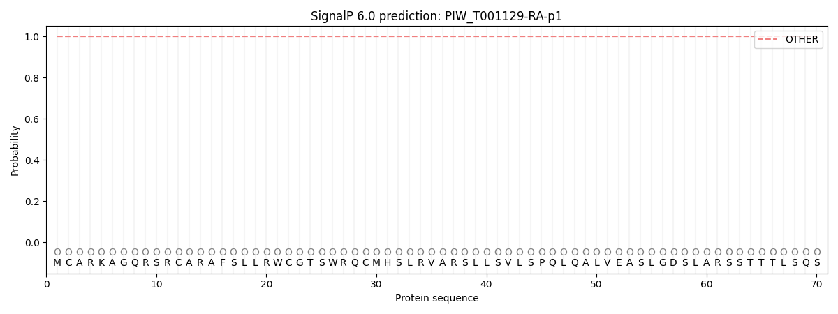
| Other | SP_Sec_SPI | CS Position |
|---|---|---|
| 1.000053 | 0.000000 |