You are browsing environment: FUNGIDB
CAZyme Information: PITG_14358-t26_1-p1
You are here: Home > Sequence: PITG_14358-t26_1-p1
Basic Information |
Genomic context |
Full Sequence |
Enzyme annotations |
CAZy signature domains |
CDD domains |
CAZyme hits |
PDB hits |
Swiss-Prot hits |
SignalP and Lipop annotations |
TMHMM annotations
Basic Information help
| Species | Phytophthora infestans | |||||||||||
|---|---|---|---|---|---|---|---|---|---|---|---|---|
| Lineage | Oomycota; NA; ; Peronosporaceae; Phytophthora; Phytophthora infestans | |||||||||||
| CAZyme ID | PITG_14358-t26_1-p1 | |||||||||||
| CAZy Family | GH81 | |||||||||||
| CAZyme Description | trans-1,2-dihydrobenzene-1,2-diol dehydrogenase, putative | |||||||||||
| CAZyme Property |
|
|||||||||||
| Genome Property |
|
|||||||||||
| Gene Location | ||||||||||||
CAZyme Signature Domains help
| Family | Start | End | Evalue | family coverage |
|---|---|---|---|---|
| GH109 | 9 | 158 | 4.4e-20 | 0.39097744360902253 |
CDD Domains download full data without filtering help
| Cdd ID | Domain | E-Value | qStart | qEnd | sStart | sEnd | Domain Description |
|---|---|---|---|---|---|---|---|
| 223745 | MviM | 2.26e-51 | 8 | 199 | 3 | 198 | Predicted dehydrogenase [General function prediction only]. |
| 396129 | GFO_IDH_MocA | 4.97e-23 | 9 | 129 | 1 | 119 | Oxidoreductase family, NAD-binding Rossmann fold. This family of enzymes utilize NADP or NAD. This family is called the GFO/IDH/MOCA family in swiss-prot. |
| 275173 | myo_inos_iolG | 1.58e-17 | 8 | 158 | 1 | 150 | inositol 2-dehydrogenase. All members of the seed alignment for this model are known or predicted inositol 2-dehydrogenase sequences co-clustered with other enzymes for catabolism of myo-inositol or closely related compounds. Inositol 2-dehydrogenase catalyzes the first step in inositol catabolism. Members of this family may vary somewhat in their ranges of acceptable substrates and some may act on analogs to myo-inositol rather than myo-inositol per se. [Energy metabolism, Sugars] |
| 182305 | PRK10206 | 4.83e-10 | 65 | 196 | 57 | 187 | putative oxidoreductase; Provisional |
| 183212 | PRK11579 | 3.66e-08 | 65 | 153 | 57 | 145 | putative oxidoreductase; Provisional |
CAZyme Hits help
| Hit ID | E-Value | Query Start | Query End | Hit Start | Hit End |
|---|---|---|---|---|---|
| 4.65e-11 | 64 | 158 | 124 | 218 | |
| 3.68e-10 | 9 | 158 | 61 | 217 | |
| 3.91e-10 | 64 | 174 | 123 | 229 | |
| 3.91e-10 | 64 | 174 | 123 | 229 | |
| 5.10e-10 | 64 | 158 | 108 | 202 |
PDB Hits download full data without filtering help
| Hit ID | E-Value | Query Start | Query End | Hit Start | Hit End | Description |
|---|---|---|---|---|---|---|
| 2.24e-41 | 7 | 207 | 1 | 202 | Crystal structure of Mammalian Dimeric Dihydrodiol Dehydrogenase [Macaca fascicularis],2O4U_X Crystal structure of Mammalian Dimeric Dihydrodiol Dehydrogenase [Macaca fascicularis],2POQ_X Dimeric Dihydrodiol Dehydrogenase complexed with inhibitor, Isoascorbic acid [Macaca fascicularis],3OHS_X Crystal Structure of Mammalian Dimeric Dihydrodiol Dehydrogenase in complex with Dihydroxyacetone [Macaca fascicularis] |
|
| 7.62e-31 | 9 | 205 | 6 | 201 | Crystal Structure of an oxidoreductase from Enterococcus faecalis [Enterococcus faecalis],3E9M_B Crystal Structure of an oxidoreductase from Enterococcus faecalis [Enterococcus faecalis],3E9M_C Crystal Structure of an oxidoreductase from Enterococcus faecalis [Enterococcus faecalis],3E9M_D Crystal Structure of an oxidoreductase from Enterococcus faecalis [Enterococcus faecalis] |
|
| 2.24e-27 | 5 | 220 | 2 | 213 | CRYSTAL STRUCTURE OF putative oxidoreductase from Streptococcus agalactiae 2603V/r [Streptococcus agalactiae serogroup V] |
|
| 2.12e-22 | 8 | 196 | 27 | 214 | Crystal Structure of KijD10, a 3-ketoreductase from Actinomadura kijaniata incomplex with NADP [Actinomadura kijaniata],3RC1_A Crystal Structure of KijD10, a 3-ketoreductase from Actinomadura kijaniata incomplex with NADP and TDP-benzene [Actinomadura kijaniata],3RC2_A Crystal Structure of KijD10, a 3-ketoreductase from Actinomadura kijaniata in complex with TDP-benzene and NADP; open conformation [Actinomadura kijaniata] |
|
| 2.93e-22 | 6 | 202 | 21 | 217 | Crystal structure of probable oxidoreductase protein from Rhizobium etli CFN 42 [Rhizobium etli CFN 42],4HAD_B Crystal structure of probable oxidoreductase protein from Rhizobium etli CFN 42 [Rhizobium etli CFN 42],4HAD_C Crystal structure of probable oxidoreductase protein from Rhizobium etli CFN 42 [Rhizobium etli CFN 42],4HAD_D Crystal structure of probable oxidoreductase protein from Rhizobium etli CFN 42 [Rhizobium etli CFN 42] |
Swiss-Prot Hits download full data without filtering help
| Hit ID | E-Value | Query Start | Query End | Hit Start | Hit End | Description |
|---|---|---|---|---|---|---|
| 1.63e-50 | 10 | 206 | 4 | 201 | Trans-1,2-dihydrobenzene-1,2-diol dehydrogenase OS=Xenopus laevis OX=8355 GN=dhdh PE=2 SV=1 |
|
| 1.63e-50 | 10 | 206 | 4 | 201 | Trans-1,2-dihydrobenzene-1,2-diol dehydrogenase OS=Xenopus tropicalis OX=8364 GN=dhdh PE=2 SV=1 |
|
| 3.42e-47 | 7 | 206 | 1 | 201 | Trans-1,2-dihydrobenzene-1,2-diol dehydrogenase OS=Danio rerio OX=7955 GN=dhdh PE=2 SV=2 |
|
| 7.11e-43 | 7 | 207 | 1 | 202 | Trans-1,2-dihydrobenzene-1,2-diol dehydrogenase OS=Bos taurus OX=9913 GN=DHDH PE=2 SV=1 |
|
| 8.70e-43 | 14 | 207 | 2 | 196 | Trans-1,2-dihydrobenzene-1,2-diol dehydrogenase (Fragment) OS=Oryctolagus cuniculus OX=9986 GN=DHDH PE=1 SV=1 |
SignalP and Lipop Annotations help
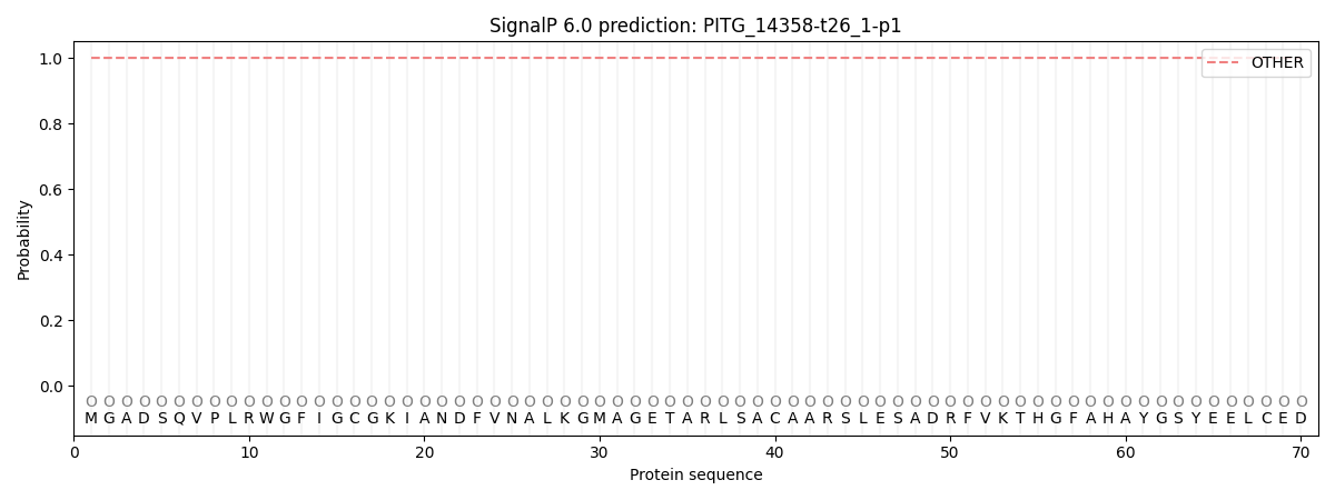
This protein is predicted as OTHER

| Other | SP_Sec_SPI | CS Position |
|---|---|---|
| 1.000077 | 0.000000 |
