You are browsing environment: FUNGIDB
CAZyme Information: PITG_06049-t26_1-p1
You are here: Home > Sequence: PITG_06049-t26_1-p1
Basic Information |
Genomic context |
Full Sequence |
Enzyme annotations |
CAZy signature domains |
CDD domains |
CAZyme hits |
PDB hits |
Swiss-Prot hits |
SignalP and Lipop annotations |
TMHMM annotations
Basic Information help
| Species | Phytophthora infestans | |||||||||||
|---|---|---|---|---|---|---|---|---|---|---|---|---|
| Lineage | Oomycota; NA; ; Peronosporaceae; Phytophthora; Phytophthora infestans | |||||||||||
| CAZyme ID | PITG_06049-t26_1-p1 | |||||||||||
| CAZy Family | GH123 | |||||||||||
| CAZyme Description | cleavage induced conserved hypothetical protein | |||||||||||
| CAZyme Property |
|
|||||||||||
| Genome Property |
|
|||||||||||
| Gene Location | ||||||||||||
CAZyme Signature Domains help
| Family | Start | End | Evalue | family coverage |
|---|---|---|---|---|
| GT8 | 147 | 387 | 1.5e-29 | 0.933852140077821 |
CDD Domains download full data without filtering help
| Cdd ID | Domain | E-Value | qStart | qEnd | sStart | sEnd | Domain Description |
|---|---|---|---|---|---|---|---|
| 133064 | GT8_GNT1 | 1.62e-38 | 162 | 393 | 1 | 245 | GNT1 is a fungal enzyme that belongs to the GT 8 family. N-acetylglucosaminyltransferase is a fungal enzyme that catalyzes the addition of N-acetyl-D-glucosamine to mannotetraose side chains by an alpha 1-2 linkage during the synthesis of mannan. The N-acetyl-D-glucosamine moiety in mannan plays a role in the attachment of mannan to asparagine residues in proteins. The mannotetraose and its N-acetyl-D-glucosamine derivative side chains of mannan are the principle immunochemical determinants on the cell surface. N-acetylglucosaminyltransferase is a member of glycosyltransferase family 8, which are, based on the relative anomeric stereochemistry of the substrate and product in the reaction catalyzed, retaining glycosyltransferases. |
| 133018 | GT8_Glycogenin | 2.23e-27 | 162 | 388 | 1 | 214 | Glycogenin belongs the GT 8 family and initiates the biosynthesis of glycogen. Glycogenin initiates the biosynthesis of glycogen by incorporating glucose residues through a self-glucosylation reaction at a Tyr residue, and then acts as substrate for chain elongation by glycogen synthase and branching enzyme. It contains a conserved DxD motif and an N-terminal beta-alpha-beta Rossmann-like fold that are common to the nucleotide-binding domains of most glycosyltransferases. The DxD motif is essential for coordination of the catalytic divalent cation, most commonly Mn2+. Glycogenin can be classified as a retaining glycosyltransferase, based on the relative anomeric stereochemistry of the substrate and product in the reaction catalyzed. It is placed in glycosyltransferase family 8 which includes lipopolysaccharide glucose and galactose transferases and galactinol synthases. |
| 227884 | Gnt1 | 3.06e-10 | 187 | 274 | 81 | 191 | Alpha-N-acetylglucosamine transferase [Cell wall/membrane/envelope biogenesis]. |
| 132996 | Glyco_transf_8 | 7.83e-09 | 223 | 338 | 66 | 208 | Members of glycosyltransferase family 8 (GT-8) are involved in lipopolysaccharide biosynthesis and glycogen synthesis. Members of this family are involved in lipopolysaccharide biosynthesis and glycogen synthesis. GT-8 comprises enzymes with a number of known activities: lipopolysaccharide galactosyltransferase, lipopolysaccharide glucosyltransferase 1, glycogenin glucosyltransferase, and N-acetylglucosaminyltransferase. GT-8 enzymes contains a conserved DXD motif which is essential in the coordination of a catalytic divalent cation, most commonly Mn2+. |
| 215090 | PLN00176 | 1.07e-08 | 241 | 386 | 103 | 266 | galactinol synthase |
CAZyme Hits help
| Hit ID | E-Value | Query Start | Query End | Hit Start | Hit End |
|---|---|---|---|---|---|
| 8.51e-39 | 222 | 422 | 176 | 371 | |
| 3.31e-29 | 135 | 387 | 40 | 299 | |
| 3.29e-27 | 157 | 386 | 123 | 365 | |
| 1.89e-26 | 163 | 398 | 10 | 263 | |
| 2.18e-26 | 139 | 406 | 56 | 339 |
PDB Hits download full data without filtering help
| Hit ID | E-Value | Query Start | Query End | Hit Start | Hit End | Description |
|---|---|---|---|---|---|---|
| 5.24e-13 | 169 | 403 | 12 | 239 | Crystal Structure of Human Glycogenin-1 (GYG1), apo form [Homo sapiens],3QVB_A Crystal Structure of Human Glycogenin-1 (GYG1) complexed with manganese and UDP [Homo sapiens],3U2W_A Crystal Structure of Human Glycogenin-1 (GYG1) complexed with manganese and glucose or a glucal species [Homo sapiens],3U2W_B Crystal Structure of Human Glycogenin-1 (GYG1) complexed with manganese and glucose or a glucal species [Homo sapiens] |
|
| 1.29e-12 | 169 | 403 | 12 | 239 | Crystal Structure of Human Glycogenin-1 (GYG1) complexed with manganese and UDP, in a triclinic closed form [Homo sapiens],3T7M_B Crystal Structure of Human Glycogenin-1 (GYG1) complexed with manganese and UDP, in a triclinic closed form [Homo sapiens],3T7N_A Crystal Structure of Human Glycogenin-1 (GYG1) complexed with manganese and UDP, in a monoclinic closed form [Homo sapiens],3T7N_B Crystal Structure of Human Glycogenin-1 (GYG1) complexed with manganese and UDP, in a monoclinic closed form [Homo sapiens],3T7O_A Crystal Structure of Human Glycogenin-1 (GYG1) complexed with manganese, UDP-Glucose and glucose [Homo sapiens],3T7O_B Crystal Structure of Human Glycogenin-1 (GYG1) complexed with manganese, UDP-Glucose and glucose [Homo sapiens],3U2U_A Crystal Structure of Human Glycogenin-1 (GYG1) complexed with manganese, UDP and maltotetraose [Homo sapiens],3U2U_B Crystal Structure of Human Glycogenin-1 (GYG1) complexed with manganese, UDP and maltotetraose [Homo sapiens],3U2V_A Crystal Structure of Human Glycogenin-1 (GYG1) complexed with manganese, UDP and maltohexaose [Homo sapiens],3U2V_B Crystal Structure of Human Glycogenin-1 (GYG1) complexed with manganese, UDP and maltohexaose [Homo sapiens],3U2X_A Crystal Structure of Human Glycogenin-1 (GYG1) complexed with manganese, UDP and 1'-deoxyglucose [Homo sapiens],3U2X_B Crystal Structure of Human Glycogenin-1 (GYG1) complexed with manganese, UDP and 1'-deoxyglucose [Homo sapiens] |
|
| 1.64e-12 | 169 | 403 | 33 | 260 | Crystal Structure of Human Glycogenin-1 (GYG1) complexed with manganese [Homo sapiens] |
|
| 1.74e-12 | 169 | 403 | 12 | 239 | Crystal Structure of Human Glycogenin-1 (GYG1) Tyr195pIPhe mutant, apo form [Homo sapiens],6EQL_A Crystal Structure of Human Glycogenin-1 (GYG1) Tyr195pIPhe mutant complexed with manganese and UDP [Homo sapiens],6EQL_B Crystal Structure of Human Glycogenin-1 (GYG1) Tyr195pIPhe mutant complexed with manganese and UDP [Homo sapiens] |
|
| 2.34e-12 | 169 | 403 | 12 | 239 | Crystal Structure of Human Glycogenin-1 (GYG1) T83M mutant complexed with manganese and UDP [Homo sapiens],3RMW_A Crystal Structure of Human Glycogenin-1 (GYG1) T83M mutant complexed with manganese and UDP-glucose [Homo sapiens] |
Swiss-Prot Hits download full data without filtering help
| Hit ID | E-Value | Query Start | Query End | Hit Start | Hit End | Description |
|---|---|---|---|---|---|---|
| 1.78e-22 | 157 | 386 | 150 | 392 | Glucose N-acetyltransferase 1 OS=Gibberella zeae (strain ATCC MYA-4620 / CBS 123657 / FGSC 9075 / NRRL 31084 / PH-1) OX=229533 GN=GNT1 PE=3 SV=2 |
|
| 8.85e-19 | 137 | 386 | 60 | 326 | Glucose N-acetyltransferase 1 OS=Neosartorya fumigata (strain ATCC MYA-4609 / Af293 / CBS 101355 / FGSC A1100) OX=330879 GN=gnt1 PE=3 SV=1 |
|
| 1.45e-11 | 169 | 403 | 11 | 238 | Glycogenin-1 OS=Homo sapiens OX=9606 GN=GYG1 PE=1 SV=4 |
|
| 3.11e-11 | 169 | 413 | 11 | 249 | Glycogenin-1 OS=Rattus norvegicus OX=10116 GN=Gyg1 PE=2 SV=4 |
|
| 5.58e-11 | 169 | 388 | 11 | 222 | Glycogenin-1 OS=Mus musculus OX=10090 GN=Gyg1 PE=1 SV=3 |
SignalP and Lipop Annotations help
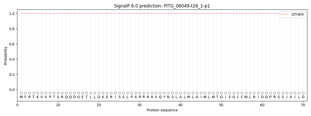
This protein is predicted as OTHER

| Other | SP_Sec_SPI | CS Position |
|---|---|---|
| 0.999984 | 0.000062 |

