You are browsing environment: FUNGIDB
CAZyme Information: PABG_11372-t30_1-p1
You are here: Home > Sequence: PABG_11372-t30_1-p1
Basic Information |
Genomic context |
Full Sequence |
Enzyme annotations |
CAZy signature domains |
CDD domains |
CAZyme hits |
PDB hits |
Swiss-Prot hits |
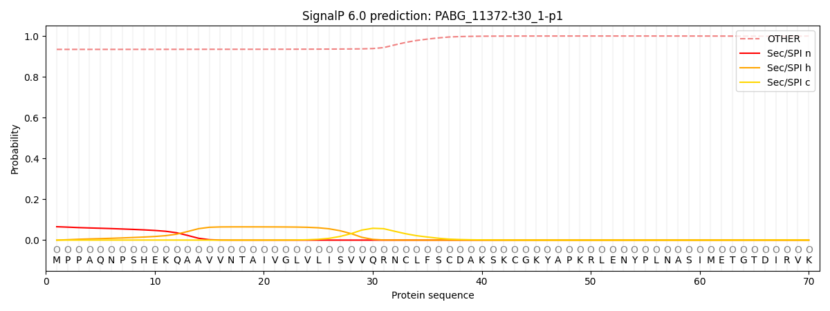
SignalP and Lipop annotations |
TMHMM annotations
Basic Information help
| Species | Paracoccidioides brasiliensis | |||||||||||
|---|---|---|---|---|---|---|---|---|---|---|---|---|
| Lineage | Ascomycota; Eurotiomycetes; ; NA; Paracoccidioides; Paracoccidioides brasiliensis | |||||||||||
| CAZyme ID | PABG_11372-t30_1-p1 | |||||||||||
| CAZy Family | GT4 | |||||||||||
| CAZyme Description | hypothetical protein | |||||||||||
| CAZyme Property |
|
|||||||||||
| Genome Property |
|
|||||||||||
| Gene Location | ||||||||||||
Enzyme Prediction help
| EC | 3.2.1.14:2 |
|---|
CAZyme Signature Domains help
| Family | Start | End | Evalue | family coverage |
|---|---|---|---|---|
| GH18 | 131 | 253 | 2.6e-25 | 0.3783783783783784 |
CDD Domains download full data without filtering help
| Cdd ID | Domain | E-Value | qStart | qEnd | sStart | sEnd | Domain Description |
|---|---|---|---|---|---|---|---|
| 119357 | GH18_zymocin_alpha | 1.01e-28 | 132 | 271 | 111 | 270 | Zymocin, alpha subunit. Zymocin is a heterotrimeric enzyme that inhibits yeast cell cycle progression. The zymocin alpha subunit has a chitinase activity that is essential for holoenzyme action from the cell exterior while the gamma subunit contains the intracellular toxin responsible for G1 phase cell cycle arrest. The zymocin alpha and beta subunits are thought to act from the cell's exterior by docking to the cell wall-associated chitin, thus mediating gamma-toxin translocation. The alpha subunit has an eight-stranded TIM barrel fold similar to that of family 18 glycosyl hydrolases such as hevamine, chitolectin, and chitobiase. |
| 214753 | Glyco_18 | 5.35e-28 | 132 | 273 | 111 | 267 | Glyco_18 domain. |
| 119365 | GH18_chitinase | 2.08e-25 | 132 | 246 | 129 | 266 | The GH18 (glycosyl hydrolases, family 18) type II chitinases hydrolyze chitin, an abundant polymer of N-acetylglucosamine and have been identified in bacteria, fungi, insects, plants, viruses, and protozoan parasites. The structure of this domain is an eight-stranded alpha/beta barrel with a pronounced active-site cleft at the C-terminal end of the beta-barrel. |
| 395573 | Glyco_hydro_18 | 9.40e-24 | 132 | 256 | 108 | 237 | Glycosyl hydrolases family 18. |
| 119351 | GH18_chitolectin_chitotriosidase | 5.09e-23 | 133 | 273 | 117 | 267 | This conserved domain family includes a large number of catalytically inactive chitinase-like lectins (chitolectins) including YKL-39, YKL-40 (HCGP39), YM1, oviductin, and AMCase (acidic mammalian chitinase), as well as catalytically active chitotriosidases. The conserved domain is an eight-stranded alpha/beta barrel fold belonging to the family 18 glycosyl hydrolases. The fold has a pronounced active-site cleft at the C-terminal end of the beta-barrel. The chitolectins lack a key active site glutamate (the proton donor required for hydrolytic activity) but retain highly conserved residues involved in oligosaccharide binding. Chitotriosidase is a chitinolytic enzyme expressed in maturing macrophages, which suggests that it plays a part in antimicrobial defense. Chitotriosidase hydrolyzes chitotriose, as well as colloidal chitin to yield chitobiose and is therefore considered an exochitinase. Chitotriosidase occurs in two major forms, the large form being converted to the small form by either RNA or post-translational processing. Although the small form, containing the chitinase domain alone, is sufficient for the chitinolytic activity, the additional C-terminal chitin-binding domain of the large form plays a role in processing colloidal chitin. The chitotriosidase gene is nonessential in humans, as about 35% of the population are heterozygous and 6% homozygous for an inactivated form of the gene. HCGP39 is a 39-kDa human cartilage glycoprotein thought to play a role in connective tissue remodeling and defense against pathogens. |
CAZyme Hits help
| Hit ID | E-Value | Query Start | Query End | Hit Start | Hit End |
|---|---|---|---|---|---|
| 6.09e-57 | 33 | 273 | 68 | 396 | |
| 1.34e-56 | 132 | 273 | 67 | 218 | |
| 4.37e-44 | 132 | 270 | 236 | 384 | |
| 1.68e-42 | 132 | 271 | 108 | 282 | |
| 2.81e-36 | 126 | 265 | 90 | 242 |
PDB Hits download full data without filtering help
| Hit ID | E-Value | Query Start | Query End | Hit Start | Hit End | Description |
|---|---|---|---|---|---|---|
| 3.03e-17 | 132 | 256 | 121 | 264 | Crystal structure of Ostrinia furnacalis Group IV chitinase [Ostrinia furnacalis],6JMB_A Chain A, ofchtiv-allosamidin [Ostrinia furnacalis] |
|
| 3.53e-16 | 132 | 256 | 121 | 264 | Crystal structure of Ostrinia furnacalis Group IV chitinase [Ostrinia furnacalis] |
|
| 9.63e-13 | 132 | 267 | 178 | 328 | Crystal structure of chitinase 40 from thermophilic bacteria Streptomyces thermoviolaceus. [Streptomyces thermoviolaceus],4W5U_B Crystal structure of chitinase 40 from thermophilic bacteria Streptomyces thermoviolaceus. [Streptomyces thermoviolaceus] |
|
| 4.86e-12 | 132 | 252 | 212 | 350 | Crystal structure of a insect group III chitinase (CAD1) from Ostrinia furnacalis [Ostrinia furnacalis],5WV9_A Crystal structure of a insect group III chitinase complex with (GlcNAc)6 (CAD1-(GlcNAc)6) from Ostrinia furnacalis [Ostrinia furnacalis] |
|
| 1.75e-11 | 132 | 276 | 289 | 475 | Crystal Structure Analysis of Chitinase A from Vibrio harveyi with novel inhibitors - apo structure of mutant W275G [Vibrio harveyi],3ART_A Crystal Structure Analysis of Chitinase A from Vibrio harveyi with novel inhibitors - W275G mutant complex structure with DEQUALINIUM [Vibrio harveyi],3ARU_A Crystal Structure Analysis of Chitinase A from Vibrio harveyi with novel inhibitors - W275G mutant complex structure with PENTOXIFYLLINE [Vibrio harveyi],3AS0_A Crystal Structure Analysis of Chitinase A from Vibrio harveyi with novel inhibitors - W275G mutant complex structure with Sanguinarine [Vibrio harveyi],3AS1_A Crystal Structure Analysis of Chitinase A from Vibrio harveyi with novel inhibitors - W275G mutant complex structure with chelerythrine [Vibrio harveyi],3AS2_A Crystal Structure Analysis of Chitinase A from Vibrio harveyi with novel inhibitors - W275G mutant complex structure with Propentofylline [Vibrio harveyi],3AS3_A Crystal Structure Analysis of Chitinase A from Vibrio harveyi with novel inhibitors - W275G mutant complex structure with 2-(imidazolin-2-yl)-5-isothiocyanatobenzofuran [Vibrio harveyi] |
Swiss-Prot Hits download full data without filtering help
| Hit ID | E-Value | Query Start | Query End | Hit Start | Hit End | Description |
|---|---|---|---|---|---|---|
| 1.73e-12 | 132 | 246 | 163 | 287 | Probable chitinase 2 OS=Drosophila melanogaster OX=7227 GN=Cht2 PE=1 SV=1 |
|
| 5.08e-11 | 132 | 277 | 378 | 541 | Chitinase 63 OS=Streptomyces plicatus OX=1922 GN=chtA PE=1 SV=2 |
|
| 1.01e-10 | 132 | 276 | 308 | 494 | Chitinase A OS=Pseudoalteromonas piscicida OX=43662 GN=chiA PE=1 SV=1 |
|
| 1.18e-10 | 132 | 249 | 141 | 277 | Endochitinase OS=Manduca sexta OX=7130 PE=2 SV=1 |
|
| 1.67e-10 | 132 | 277 | 377 | 540 | Chitinase C OS=Streptomyces lividans OX=1916 GN=chiC PE=2 SV=1 |
SignalP and Lipop Annotations help
This protein is predicted as OTHER

| Other | SP_Sec_SPI | CS Position |
|---|---|---|
| 0.938471 | 0.061563 |
