You are browsing environment: FUNGIDB
CAZyme Information: OAG18044.1
You are here: Home > Sequence: OAG18044.1
Basic Information |
Genomic context |
Full Sequence |
Enzyme annotations |
CAZy signature domains |
CDD domains |
CAZyme hits |
PDB hits |
Swiss-Prot hits |
SignalP and Lipop annotations |
TMHMM annotations
Basic Information help
| Species | Alternaria alternata | |||||||||||
|---|---|---|---|---|---|---|---|---|---|---|---|---|
| Lineage | Ascomycota; Dothideomycetes; ; Pleosporaceae; Alternaria; Alternaria alternata | |||||||||||
| CAZyme ID | OAG18044.1 | |||||||||||
| CAZy Family | GH105 | |||||||||||
| CAZyme Description | glycosyl transferas-like protein | |||||||||||
| CAZyme Property |
|
|||||||||||
| Genome Property |
|
|||||||||||
| Gene Location | ||||||||||||
CAZyme Signature Domains help
| Family | Start | End | Evalue | family coverage |
|---|---|---|---|---|
| GT1 | 64 | 431 | 1.1e-30 | 0.8481675392670157 |
CDD Domains download full data without filtering help
| Cdd ID | Domain | E-Value | qStart | qEnd | sStart | sEnd | Domain Description |
|---|---|---|---|---|---|---|---|
| 224732 | YjiC | 1.16e-27 | 9 | 432 | 4 | 399 | UDP:flavonoid glycosyltransferase YjiC, YdhE family [Carbohydrate transport and metabolism]. |
| 340817 | GT1_Gtf-like | 5.21e-26 | 7 | 427 | 2 | 400 | UDP-glycosyltransferases and similar proteins. This family includes the Gtfs, a group of homologous glycosyltransferases involved in the final stages of the biosynthesis of antibiotics vancomycin and related chloroeremomycin. Gtfs transfer sugar moieties from an activated NDP-sugar donor to the oxidatively cross-linked heptapeptide core of vancomycin group antibiotics. The core structure is important for the bioactivity of the antibiotics. |
| 223779 | MurG | 3.14e-10 | 98 | 428 | 79 | 351 | UDP-N-acetylglucosamine:LPS N-acetylglucosamine transferase [Cell wall/membrane/envelope biogenesis]. |
| 340818 | GT28_MurG | 2.43e-08 | 17 | 427 | 10 | 350 | undecaprenyldiphospho-muramoylpentapeptide beta-N-acetylglucosaminyltransferase. MurG (EC 2.4.1.227) is an N-acetylglucosaminyltransferase, the last enzyme involved in the intracellular phase of peptidoglycan biosynthesis. It transfers N-acetyl-D-glucosamine (GlcNAc) from UDP-GlcNAc to the C4 hydroxyl of a lipid-linked N-acetylmuramoyl pentapeptide (NAM). The resulting disaccharide is then transported across the cell membrane, where it is polymerized into NAG-NAM cell-wall repeat structure. MurG belongs to the GT-B structural superfamily of glycoslytransferases, which have characteristic N- and C-terminal domains, each containing a typical Rossmann fold. The two domains have high structural homology despite minimal sequence homology. The large cleft that separates the two domains includes the catalytic center and permits a high degree of flexibility. |
| 397977 | Glyco_tran_28_C | 4.08e-04 | 332 | 412 | 70 | 155 | Glycosyltransferase family 28 C-terminal domain. The glycosyltransferase family 28 includes monogalactosyldiacylglycerol synthase (EC 2.4.1.46) and UDP-N-acetylglucosamine transferase (EC 2.4.1.-). Structural analysis suggests the C-terminal domain contains the UDP-GlcNAc binding site. |
CAZyme Hits help
| Hit ID | E-Value | Query Start | Query End | Hit Start | Hit End |
|---|---|---|---|---|---|
| 3.75e-298 | 1 | 439 | 1 | 439 | |
| 1.77e-297 | 1 | 445 | 1 | 443 | |
| 2.43e-287 | 1 | 440 | 1 | 440 | |
| 8.40e-282 | 26 | 439 | 1 | 414 | |
| 5.23e-104 | 4 | 435 | 5 | 440 |
PDB Hits download full data without filtering help
| Hit ID | E-Value | Query Start | Query End | Hit Start | Hit End | Description |
|---|---|---|---|---|---|---|
| 6.95e-10 | 24 | 428 | 7 | 390 | Crystal Structure Analysis of the Glycotransferase of kitacinnamycin [Kitasatospora],6J31_B Crystal Structure Analysis of the Glycotransferase of kitacinnamycin [Kitasatospora],6J31_C Crystal Structure Analysis of the Glycotransferase of kitacinnamycin [Kitasatospora],6J31_D Crystal Structure Analysis of the Glycotransferase of kitacinnamycin [Kitasatospora] |
|
| 7.02e-10 | 24 | 428 | 10 | 393 | Crystal Structure Analysis of the Glycotransferase of kitacinnamycin [Kitasatospora],6J32_B Crystal Structure Analysis of the Glycotransferase of kitacinnamycin [Kitasatospora],6J32_C Crystal Structure Analysis of the Glycotransferase of kitacinnamycin [Kitasatospora],6J32_D Crystal Structure Analysis of the Glycotransferase of kitacinnamycin [Kitasatospora] |
|
| 2.82e-07 | 262 | 420 | 243 | 395 | Crystal Structure of CalG1, Calicheamicin Glycostyltransferase, TDP bound form [Micromonospora echinospora],3OTH_A Crystal Structure of CalG1, Calicheamicin Glycostyltransferase, TDP and calicheamicin alpha3I bound form [Micromonospora echinospora],3OTH_B Crystal Structure of CalG1, Calicheamicin Glycostyltransferase, TDP and calicheamicin alpha3I bound form [Micromonospora echinospora] |
|
| 9.14e-07 | 322 | 419 | 322 | 419 | Structural Characterization of UDP-glycosyltransferase from Tetranychus Urticae [Tetranychus urticae] |
|
| 9.82e-07 | 338 | 412 | 91 | 165 | Crystal Structure of the UDP-Glucuronic Acid Binding Domain of the Human Drug Metabolizing UDP-Glucuronosyltransferase 2B7 [Homo sapiens],2O6L_B Crystal Structure of the UDP-Glucuronic Acid Binding Domain of the Human Drug Metabolizing UDP-Glucuronosyltransferase 2B7 [Homo sapiens] |
Swiss-Prot Hits download full data without filtering help
| Hit ID | E-Value | Query Start | Query End | Hit Start | Hit End | Description |
|---|---|---|---|---|---|---|
| 2.56e-14 | 93 | 413 | 120 | 418 | PGL/p-HBAD biosynthesis rhamnosyltransferase OS=Mycobacterium bovis (strain BCG / Pasteur 1173P2) OX=410289 GN=BCG_2983c PE=3 SV=1 |
|
| 2.56e-14 | 93 | 413 | 120 | 418 | PGL/p-HBAD biosynthesis rhamnosyltransferase OS=Mycobacterium tuberculosis (strain CDC 1551 / Oshkosh) OX=83331 GN=MT3038 PE=3 SV=1 |
|
| 2.56e-14 | 93 | 413 | 120 | 418 | PGL/p-HBAD biosynthesis rhamnosyltransferase OS=Mycobacterium tuberculosis (strain ATCC 25618 / H37Rv) OX=83332 GN=Rv2962c PE=1 SV=1 |
|
| 2.56e-14 | 93 | 413 | 120 | 418 | PGL/p-HBAD biosynthesis rhamnosyltransferase OS=Mycobacterium tuberculosis (strain ATCC 25177 / H37Ra) OX=419947 GN=MRA_2989 PE=3 SV=1 |
|
| 2.56e-14 | 93 | 413 | 120 | 418 | PGL/p-HBAD biosynthesis rhamnosyltransferase OS=Mycobacterium bovis (strain ATCC BAA-935 / AF2122/97) OX=233413 GN=BQ2027_MB2986C PE=3 SV=1 |
SignalP and Lipop Annotations help
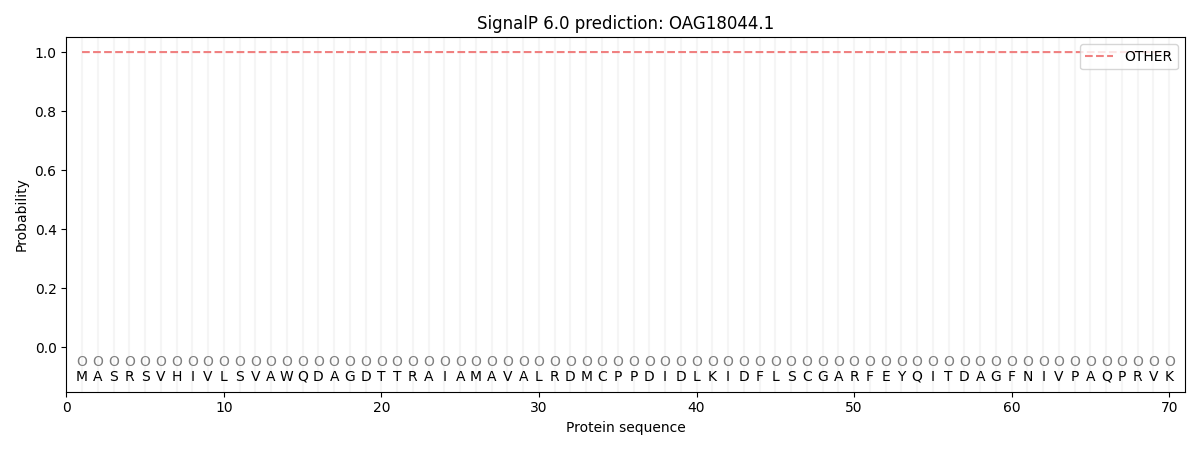
This protein is predicted as OTHER

| Other | SP_Sec_SPI | CS Position |
|---|---|---|
| 1.000101 | 0.000000 |
