You are browsing environment: FUNGIDB
CAZyme Information: KIW98341.1
You are here: Home > Sequence: KIW98341.1
Basic Information |
Genomic context |
Full Sequence |
Enzyme annotations |
CAZy signature domains |
CDD domains |
CAZyme hits |
PDB hits |
Swiss-Prot hits |
SignalP and Lipop annotations |
TMHMM annotations
Basic Information help
| Species | Cladophialophora bantiana | |||||||||||
|---|---|---|---|---|---|---|---|---|---|---|---|---|
| Lineage | Ascomycota; Eurotiomycetes; ; Herpotrichiellaceae; Cladophialophora; Cladophialophora bantiana | |||||||||||
| CAZyme ID | KIW98341.1 | |||||||||||
| CAZy Family | GT48 | |||||||||||
| CAZyme Description | hypothetical protein | |||||||||||
| CAZyme Property |
|
|||||||||||
| Genome Property |
|
|||||||||||
| Gene Location | ||||||||||||
CAZyme Signature Domains help
| Family | Start | End | Evalue | family coverage |
|---|---|---|---|---|
| AA4 | 35 | 576 | 1e-147 | 0.9885057471264368 |
CDD Domains download full data without filtering help
| Cdd ID | Domain | E-Value | qStart | qEnd | sStart | sEnd | Domain Description |
|---|---|---|---|---|---|---|---|
| 223354 | GlcD | 3.10e-30 | 52 | 567 | 1 | 455 | FAD/FMN-containing dehydrogenase [Energy production and conversion]. |
| 396238 | FAD_binding_4 | 4.98e-30 | 87 | 234 | 1 | 139 | FAD binding domain. This family consists of various enzymes that use FAD as a co-factor, most of the enzymes are similar to oxygen oxidoreductase. One of the enzymes Vanillyl-alcohol oxidase (VAO) has a solved structure, the alignment includes the FAD binding site, called the PP-loop, between residues 99-110. The FAD molecule is covalently bound in the known structure, however the residue that links to the FAD is not in the alignment. VAO catalyzes the oxidation of a wide variety of substrates, ranging form aromatic amines to 4-alkylphenols. Other members of this family include D-lactate dehydrogenase, this enzyme catalyzes the conversion of D-lactate to pyruvate using FAD as a co-factor; mitomycin radical oxidase, this enzyme oxidizes the reduced form of mitomycins and is involved in mitomycin resistance. This family includes MurB an UDP-N-acetylenolpyruvoylglucosamine reductase enzyme EC:1.1.1.158. This enzyme is involved in the biosynthesis of peptidoglycan. |
| 183043 | PRK11230 | 1.24e-10 | 39 | 295 | 14 | 245 | glycolate oxidase subunit GlcD; Provisional |
| 178402 | PLN02805 | 1.83e-09 | 86 | 293 | 133 | 323 | D-lactate dehydrogenase [cytochrome] |
| 273751 | FAD_lactone_ox | 1.49e-05 | 87 | 231 | 15 | 147 | sugar 1,4-lactone oxidases. This model represents a family of at least two different sugar 1,4 lactone oxidases, both involved in synthesizing ascorbic acid or a derivative. These include L-gulonolactone oxidase (EC 1.1.3.8) from rat and D-arabinono-1,4-lactone oxidase (EC 1.1.3.37) from Saccharomyces cerevisiae. Members are proposed to have the cofactor FAD covalently bound at a site specified by Prosite motif PS00862; OX2_COVAL_FAD; 1. |
CAZyme Hits help
| Hit ID | E-Value | Query Start | Query End | Hit Start | Hit End |
|---|---|---|---|---|---|
| 8.27e-300 | 6 | 575 | 1 | 560 | |
| 9.25e-299 | 12 | 575 | 6 | 559 | |
| 2.94e-287 | 6 | 581 | 4 | 570 | |
| 1.44e-280 | 6 | 580 | 4 | 569 | |
| 4.67e-249 | 12 | 580 | 10 | 568 |
PDB Hits download full data without filtering help
| Hit ID | E-Value | Query Start | Query End | Hit Start | Hit End | Description |
|---|---|---|---|---|---|---|
| 3.01e-109 | 33 | 575 | 1 | 522 | Crystal structure of eugenol oxidase in complex with isoeugenol [Rhodococcus jostii RHA1],5FXD_B Crystal structure of eugenol oxidase in complex with isoeugenol [Rhodococcus jostii RHA1],5FXE_A Crystal structure of eugenol oxidase in complex with coniferyl alcohol [Rhodococcus jostii RHA1],5FXE_B Crystal structure of eugenol oxidase in complex with coniferyl alcohol [Rhodococcus jostii RHA1],5FXF_A Crystal structure of eugenol oxidase in complex with benzoate [Rhodococcus jostii RHA1],5FXF_B Crystal structure of eugenol oxidase in complex with benzoate [Rhodococcus jostii RHA1],5FXP_A Crystal structure of eugenol oxidase in complex with vanillin [Rhodococcus jostii RHA1],5FXP_B Crystal structure of eugenol oxidase in complex with vanillin [Rhodococcus jostii RHA1] |
|
| 6.51e-104 | 37 | 581 | 12 | 560 | Structure of the D170S/T457E double mutant of vanillyl-alcohol oxidase [Penicillium simplicissimum],1E0Y_B Structure of the D170S/T457E double mutant of vanillyl-alcohol oxidase [Penicillium simplicissimum] |
|
| 9.16e-104 | 37 | 581 | 12 | 560 | STRUCTURE OF THE OCTAMERIC FLAVOENZYME VANILLYL-ALCOHOL OXIDASE: The505Ser Mutant [Penicillium simplicissimum],1W1J_B STRUCTURE OF THE OCTAMERIC FLAVOENZYME VANILLYL-ALCOHOL OXIDASE: The505Ser Mutant [Penicillium simplicissimum] |
|
| 9.16e-104 | 37 | 581 | 12 | 560 | STRUCTURE OF THE OCTAMERIC FLAVOENZYME VANILLYL-ALCOHOL OXIDASE: Ile238Thr Mutant [Penicillium simplicissimum],1W1K_B STRUCTURE OF THE OCTAMERIC FLAVOENZYME VANILLYL-ALCOHOL OXIDASE: Ile238Thr Mutant [Penicillium simplicissimum] |
|
| 1.81e-103 | 37 | 581 | 12 | 560 | Asp170Ser mutant of vanillyl-alcohol oxidase [Penicillium simplicissimum],1DZN_B Asp170Ser mutant of vanillyl-alcohol oxidase [Penicillium simplicissimum] |
Swiss-Prot Hits download full data without filtering help
| Hit ID | E-Value | Query Start | Query End | Hit Start | Hit End | Description |
|---|---|---|---|---|---|---|
| 1.31e-102 | 37 | 581 | 12 | 560 | Vanillyl-alcohol oxidase OS=Penicillium simplicissimum OX=69488 GN=VAOA PE=1 SV=1 |
|
| 2.48e-90 | 36 | 569 | 8 | 515 | 4-cresol dehydrogenase [hydroxylating] flavoprotein subunit OS=Pseudomonas putida OX=303 GN=pchF PE=1 SV=3 |
|
| 1.60e-20 | 53 | 318 | 8 | 253 | Uncharacterized FAD-linked oxidoreductase Rv2280 OS=Mycobacterium tuberculosis (strain ATCC 25618 / H37Rv) OX=83332 GN=Rv2280 PE=1 SV=1 |
|
| 1.60e-20 | 53 | 318 | 8 | 253 | Uncharacterized FAD-linked oxidoreductase MT2338 OS=Mycobacterium tuberculosis (strain CDC 1551 / Oshkosh) OX=83331 GN=MT2338 PE=3 SV=1 |
|
| 1.73e-16 | 84 | 314 | 38 | 250 | Glycolate oxidase subunit GlcD OS=Bacillus subtilis (strain 168) OX=224308 GN=glcD PE=3 SV=1 |
SignalP and Lipop Annotations help
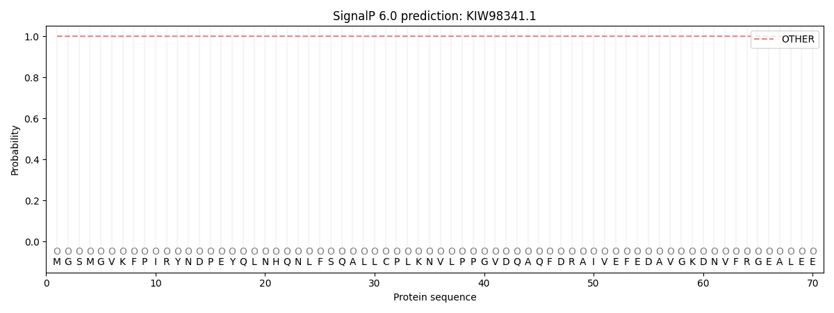
This protein is predicted as OTHER

| Other | SP_Sec_SPI | CS Position |
|---|---|---|
| 1.000046 | 0.000000 |
