You are browsing environment: FUNGIDB
CAZyme Information: KID68755.1
You are here: Home > Sequence: KID68755.1
Basic Information |
Genomic context |
Full Sequence |
Enzyme annotations |
CAZy signature domains |
CDD domains |
CAZyme hits |
PDB hits |
Swiss-Prot hits |
SignalP and Lipop annotations |
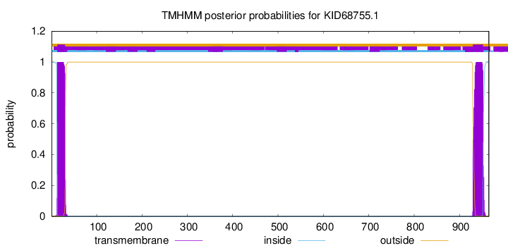
TMHMM annotations
Basic Information help
| Species | Metarhizium anisopliae | |||||||||||
|---|---|---|---|---|---|---|---|---|---|---|---|---|
| Lineage | Ascomycota; Sordariomycetes; ; Clavicipitaceae; Metarhizium; Metarhizium anisopliae | |||||||||||
| CAZyme ID | KID68755.1 | |||||||||||
| CAZy Family | GH79 | |||||||||||
| CAZyme Description | Glycoside hydrolase, family 47 | |||||||||||
| CAZyme Property |
|
|||||||||||
| Genome Property |
|
|||||||||||
| Gene Location | ||||||||||||
Enzyme Prediction help
| EC | 3.2.1.113:7 |
|---|
CAZyme Signature Domains help
| Family | Start | End | Evalue | family coverage |
|---|---|---|---|---|
| GH47 | 103 | 566 | 8.2e-154 | 0.9977578475336323 |
CDD Domains download full data without filtering help
| Cdd ID | Domain | E-Value | qStart | qEnd | sStart | sEnd | Domain Description |
|---|---|---|---|---|---|---|---|
| 396217 | Glyco_hydro_47 | 0.0 | 103 | 566 | 1 | 453 | Glycosyl hydrolase family 47. Members of this family are alpha-mannosidases that catalyze the hydrolysis of the terminal 1,2-linked alpha-D-mannose residues in the oligo-mannose oligosaccharide Man(9)(GlcNAc)(2). |
| 240427 | PTZ00470 | 4.79e-119 | 88 | 567 | 64 | 519 | glycoside hydrolase family 47 protein; Provisional |
| 395170 | DnaJ | 1.93e-17 | 678 | 769 | 1 | 63 | DnaJ domain. DnaJ domains (J-domains) are associated with hsp70 heat-shock system and it is thought that this domain mediates the interaction. DnaJ-domain is therefore part of a chaperone (protein folding) system. The T-antigens, although not in Prosite are confirmed as DnaJ containing domains from literature. |
| 237656 | PRK14280 | 1.69e-14 | 675 | 770 | 2 | 67 | molecular chaperone DnaJ. |
| 274090 | DnaJ_bact | 3.33e-14 | 680 | 770 | 3 | 63 | chaperone protein DnaJ. This model represents bacterial forms of DnaJ, part of the DnaK-DnaJ-GrpE chaperone system. The three components typically are encoded by consecutive genes. DnaJ homologs occur in many genomes, typically not near DnaK and GrpE-like genes; most such genes are not included by this family. Eukaryotic (mitochondrial and chloroplast) forms are not included in the scope of this family. |
CAZyme Hits help
| Hit ID | E-Value | Query Start | Query End | Hit Start | Hit End |
|---|---|---|---|---|---|
| 0.0 | 1 | 570 | 1 | 570 | |
| 0.0 | 1 | 570 | 1 | 579 | |
| 3.57e-316 | 9 | 571 | 9 | 574 | |
| 4.44e-291 | 3 | 963 | 4 | 814 | |
| 4.31e-284 | 3 | 965 | 4 | 869 |
PDB Hits download full data without filtering help
| Hit ID | E-Value | Query Start | Query End | Hit Start | Hit End | Description |
|---|---|---|---|---|---|---|
| 6.02e-69 | 92 | 564 | 1 | 471 | Penicillium citrinum alpha-1,2-mannosidase complex with glycerol [Penicillium citrinum],2RI8_B Penicillium citrinum alpha-1,2-mannosidase complex with glycerol [Penicillium citrinum],2RI9_A Penicillium citrinum alpha-1,2-mannosidase in complex with a substrate analog [Penicillium citrinum],2RI9_B Penicillium citrinum alpha-1,2-mannosidase in complex with a substrate analog [Penicillium citrinum] |
|
| 7.81e-69 | 78 | 564 | 22 | 506 | Structure of P. citrinum alpha 1,2-mannosidase reveals the basis for differences in specificity of the ER and Golgi Class I enzymes [Penicillium citrinum],1KKT_B Structure of P. citrinum alpha 1,2-mannosidase reveals the basis for differences in specificity of the ER and Golgi Class I enzymes [Penicillium citrinum],1KRE_A Structure Of P. Citrinum Alpha 1,2-Mannosidase Reveals The Basis For Differences In Specificity Of The Er And Golgi Class I Enzymes [Penicillium citrinum],1KRE_B Structure Of P. Citrinum Alpha 1,2-Mannosidase Reveals The Basis For Differences In Specificity Of The Er And Golgi Class I Enzymes [Penicillium citrinum],1KRF_A Structure Of P. Citrinum Alpha 1,2-mannosidase Reveals The Basis For Differences In Specificity Of The Er And Golgi Class I Enzymes [Penicillium citrinum],1KRF_B Structure Of P. Citrinum Alpha 1,2-mannosidase Reveals The Basis For Differences In Specificity Of The Er And Golgi Class I Enzymes [Penicillium citrinum] |
|
| 9.81e-64 | 50 | 564 | 43 | 532 | Crystal Structure Of Human Class I alpha-1,2-Mannosidase In Complex With Thio-Disaccharide Substrate Analogue [Homo sapiens] |
|
| 1.16e-63 | 96 | 564 | 5 | 449 | Crystal structure of the class I human endoplasmic reticulum 1,2-alpha-mannosidase and Man9GlcNAc2-PA complex [Homo sapiens] |
|
| 1.29e-63 | 94 | 563 | 20 | 464 | Structure of mouse Golgi alpha-1,2-mannosidase IA and Man9GlcNAc2-PA complex [Mus musculus],5KKB_B Structure of mouse Golgi alpha-1,2-mannosidase IA and Man9GlcNAc2-PA complex [Mus musculus] |
Swiss-Prot Hits download full data without filtering help
| Hit ID | E-Value | Query Start | Query End | Hit Start | Hit End | Description |
|---|---|---|---|---|---|---|
| 6.30e-73 | 93 | 565 | 30 | 500 | Mannosyl-oligosaccharide alpha-1,2-mannosidase 1B OS=Emericella nidulans (strain FGSC A4 / ATCC 38163 / CBS 112.46 / NRRL 194 / M139) OX=227321 GN=mns1B PE=2 SV=2 |
|
| 1.47e-72 | 89 | 565 | 35 | 509 | Probable mannosyl-oligosaccharide alpha-1,2-mannosidase 1B OS=Aspergillus niger (strain CBS 513.88 / FGSC A1513) OX=425011 GN=mns1B PE=3 SV=1 |
|
| 8.93e-71 | 91 | 565 | 35 | 507 | Probable mannosyl-oligosaccharide alpha-1,2-mannosidase 1B OS=Aspergillus flavus (strain ATCC 200026 / FGSC A1120 / IAM 13836 / NRRL 3357 / JCM 12722 / SRRC 167) OX=332952 GN=mns1B PE=3 SV=2 |
|
| 8.93e-71 | 91 | 565 | 35 | 507 | Mannosyl-oligosaccharide alpha-1,2-mannosidase 1B OS=Aspergillus oryzae (strain ATCC 42149 / RIB 40) OX=510516 GN=mns1B PE=1 SV=1 |
|
| 9.62e-71 | 91 | 565 | 37 | 509 | Mannosyl-oligosaccharide alpha-1,2-mannosidase 1B OS=Aspergillus phoenicis OX=5063 GN=mns1B PE=2 SV=1 |
SignalP and Lipop Annotations help
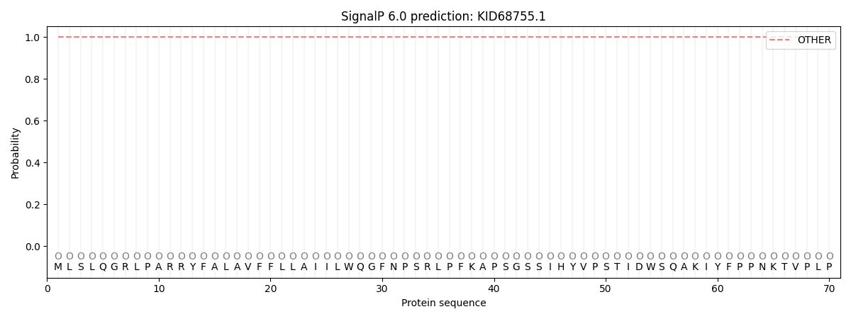
This protein is predicted as OTHER

| Other | SP_Sec_SPI | CS Position |
|---|---|---|
| 0.999995 | 0.000002 |

