You are browsing environment: FUNGIDB
CAZyme Information: KAG1708657.1
You are here: Home > Sequence: KAG1708657.1
Basic Information |
Genomic context |
Full Sequence |
Enzyme annotations |
CAZy signature domains |
CDD domains |
CAZyme hits |
PDB hits |
Swiss-Prot hits |
SignalP and Lipop annotations |
TMHMM annotations
Basic Information help
| Species | Phytophthora capsici | |||||||||||
|---|---|---|---|---|---|---|---|---|---|---|---|---|
| Lineage | Oomycota; NA; ; Peronosporaceae; Phytophthora; Phytophthora capsici | |||||||||||
| CAZyme ID | KAG1708657.1 | |||||||||||
| CAZy Family | GT41 | |||||||||||
| CAZyme Description | unspecified product | |||||||||||
| CAZyme Property |
|
|||||||||||
| Genome Property |
|
|||||||||||
| Gene Location | ||||||||||||
CAZyme Signature Domains help
| Family | Start | End | Evalue | family coverage |
|---|---|---|---|---|
| CBM47 | 1114 | 1254 | 8.3e-19 | 0.96875 |
CDD Domains download full data without filtering help
| Cdd ID | Domain | E-Value | qStart | qEnd | sStart | sEnd | Domain Description |
|---|---|---|---|---|---|---|---|
| 227511 | ATS1 | 1.55e-28 | 806 | 1082 | 94 | 374 | Alpha-tubulin suppressor and related RCC1 domain-containing proteins [Cell cycle control, cell division, chromosome partitioning, Cytoskeleton]. |
| 227455 | FRQ1 | 9.64e-21 | 82 | 234 | 7 | 156 | Ca2+-binding protein, EF-hand superfamily [Signal transduction mechanisms]. |
| 227511 | ATS1 | 1.36e-18 | 828 | 1085 | 59 | 321 | Alpha-tubulin suppressor and related RCC1 domain-containing proteins [Cell cycle control, cell division, chromosome partitioning, Cytoskeleton]. |
| 227511 | ATS1 | 2.04e-17 | 809 | 982 | 275 | 440 | Alpha-tubulin suppressor and related RCC1 domain-containing proteins [Cell cycle control, cell division, chromosome partitioning, Cytoskeleton]. |
| 320055 | EFh_PEF_Group_I | 1.12e-16 | 126 | 230 | 1 | 91 | Penta-EF hand, calcium binding motifs, found in Group I PEF proteins. The family corresponds to Group I PEF proteins that have been found not only in higher animals but also in lower animals, plants, fungi and protists. Group I PEF proteins include apoptosis-linked gene 2 protein (ALG-2), peflin and similar proteins. ALG-2, also termed programmed cell death protein 6 (PDCD6), is a widely expressed calcium-binding modulator protein associated with cell proliferation and death, as well as cell survival. It forms a homodimer in the cell or a heterodimer with its closest paralog peflin. Among the PEF proteins, ALG-2 can bind three Ca2+ ions through its EF1, EF3, and EF5 hands, where it is unique in that its EF5 hand binds Ca2+ ion in a canonical coordination. Peflin is a ubiquitously expressed 30-kD PEF protein containing five EF-hand motifs in its C-terminal domain and a longer N-terminal hydrophobic domain (NHB domain) than any other member of the PEF family. The NHB domain harbors nine repeats of a nonapeptide (A/PPGGPYGGP). Peflin may modulate the function of ALG-2 in Ca2+ signaling. It exists only as a heterodimer with ALG-2, and binds two Ca2+ ions through its EF1 and EF3 hands. Its additional EF5 hand is unpaired and does not bind Ca2+ ion but mediates the heterodimerization with ALG-2. The dissociation of heterodimer occurs in the presence of Ca2+. |
CAZyme Hits help
| Hit ID | E-Value | Query Start | Query End | Hit Start | Hit End |
|---|---|---|---|---|---|
| 2.82e-14 | 820 | 1078 | 93 | 328 | |
| 1.10e-13 | 820 | 1078 | 91 | 326 | |
| 1.10e-12 | 1104 | 1258 | 203 | 340 | |
| 1.10e-12 | 1104 | 1258 | 203 | 340 | |
| 1.19e-11 | 1040 | 1256 | 653 | 856 |
PDB Hits download full data without filtering help
| Hit ID | E-Value | Query Start | Query End | Hit Start | Hit End | Description |
|---|---|---|---|---|---|---|
| 6.95e-25 | 818 | 1101 | 109 | 369 | Crystal structure of the W285F mutant of UVB-resistance protein UVR8 [Arabidopsis thaliana],4DNV_B Crystal structure of the W285F mutant of UVB-resistance protein UVR8 [Arabidopsis thaliana],4DNV_C Crystal structure of the W285F mutant of UVB-resistance protein UVR8 [Arabidopsis thaliana],4DNV_D Crystal structure of the W285F mutant of UVB-resistance protein UVR8 [Arabidopsis thaliana] |
|
| 7.29e-25 | 818 | 1101 | 112 | 372 | Chain A, Ultraviolet-B receptor UVR8 [Arabidopsis thaliana],6XZM_B Chain B, Ultraviolet-B receptor UVR8 [Arabidopsis thaliana] |
|
| 7.29e-25 | 818 | 1101 | 112 | 372 | Chain A, Ultraviolet-B receptor UVR8 [Arabidopsis thaliana],6XZN_B Chain B, Ultraviolet-B receptor UVR8 [Arabidopsis thaliana] |
|
| 7.78e-25 | 818 | 1101 | 108 | 368 | Crystal structure of photoreceptor AtUVR8 mutant W285F and light-induced structural changes at 120K [Arabidopsis thaliana],4NC4_B Crystal structure of photoreceptor AtUVR8 mutant W285F and light-induced structural changes at 120K [Arabidopsis thaliana],4NC4_C Crystal structure of photoreceptor AtUVR8 mutant W285F and light-induced structural changes at 120K [Arabidopsis thaliana],4NC4_D Crystal structure of photoreceptor AtUVR8 mutant W285F and light-induced structural changes at 120K [Arabidopsis thaliana] |
|
| 9.98e-25 | 818 | 1101 | 109 | 369 | Crystal structure of UVB-resistance protein UVR8 [Arabidopsis thaliana],4DNW_B Crystal structure of UVB-resistance protein UVR8 [Arabidopsis thaliana] |
Swiss-Prot Hits download full data without filtering help
| Hit ID | E-Value | Query Start | Query End | Hit Start | Hit End | Description |
|---|---|---|---|---|---|---|
| 7.50e-27 | 822 | 1093 | 3023 | 3275 | Probable E3 ubiquitin-protein ligase HERC2 OS=Drosophila melanogaster OX=7227 GN=HERC2 PE=1 SV=3 |
|
| 8.88e-27 | 824 | 1082 | 29 | 270 | Probable E3 ubiquitin-protein ligase HERC6 OS=Homo sapiens OX=9606 GN=HERC6 PE=1 SV=2 |
|
| 4.27e-25 | 801 | 1174 | 2976 | 3351 | E3 ubiquitin-protein ligase HERC2 OS=Mus musculus OX=10090 GN=Herc2 PE=1 SV=3 |
|
| 5.58e-25 | 801 | 1174 | 2975 | 3350 | E3 ubiquitin-protein ligase HERC2 OS=Homo sapiens OX=9606 GN=HERC2 PE=1 SV=2 |
|
| 1.30e-23 | 818 | 1101 | 120 | 380 | Ultraviolet-B receptor UVR8 OS=Arabidopsis thaliana OX=3702 GN=UVR8 PE=1 SV=1 |
SignalP and Lipop Annotations help
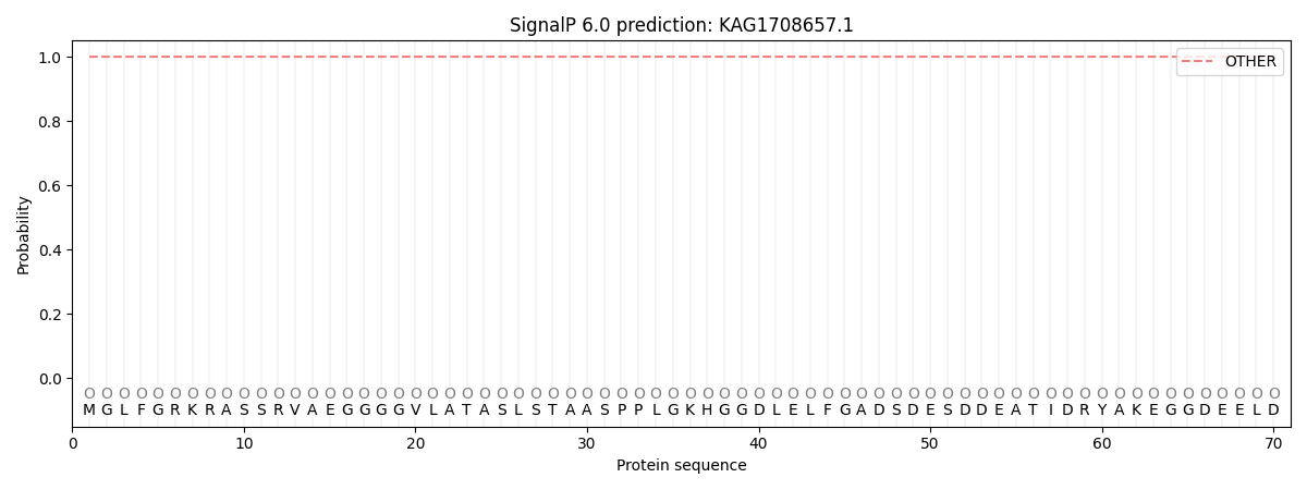
This protein is predicted as OTHER

| Other | SP_Sec_SPI | CS Position |
|---|---|---|
| 1.000042 | 0.000014 |
