You are browsing environment: FUNGIDB
CAZyme Information: I306_02179-t38_1-p1
You are here: Home > Sequence: I306_02179-t38_1-p1
Basic Information |
Genomic context |
Full Sequence |
Enzyme annotations |
CAZy signature domains |
CDD domains |
CAZyme hits |
PDB hits |
Swiss-Prot hits |
SignalP and Lipop annotations |
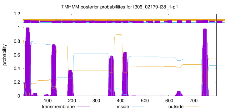
TMHMM annotations
Basic Information help
| Species | Cryptococcus gattii VGI | |||||||||||
|---|---|---|---|---|---|---|---|---|---|---|---|---|
| Lineage | Arthropoda; Insecta; ; Eriococcidae; Cryptococcus; Cryptococcus gattii VGI | |||||||||||
| CAZyme ID | I306_02179-t38_1-p1 | |||||||||||
| CAZy Family | GH13 | |||||||||||
| CAZyme Description | phosphatidylinositol glycan, class A | |||||||||||
| CAZyme Property |
|
|||||||||||
| Genome Property |
|
|||||||||||
| Gene Location | ||||||||||||
Enzyme Prediction help
| EC | 2.4.1.198:3 |
|---|
CDD Domains download full data without filtering help
| Cdd ID | Domain | E-Value | qStart | qEnd | sStart | sEnd | Domain Description |
|---|---|---|---|---|---|---|---|
| 340827 | GT4_PIG-A-like | 0.0 | 355 | 751 | 2 | 398 | phosphatidylinositol N-acetylglucosaminyltransferase subunit A and similar proteins. This family is most closely related to the GT4 family of glycosyltransferases. Phosphatidylinositol glycan-class A (PIG-A), an X-linked gene in humans, is necessary for the synthesis of N-acetylglucosaminyl-phosphatidylinositol, a very early intermediate in glycosyl phosphatidylinositol (GPI)-anchor biosynthesis. The GPI-anchor is an important cellular structure that facilitates the attachment of many proteins to cell surfaces. Somatic mutations in PIG-A have been associated with Paroxysmal Nocturnal Hemoglobinuria (PNH), an acquired hematological disorder. |
| 340831 | GT4_PimA-like | 8.58e-55 | 355 | 718 | 2 | 365 | phosphatidyl-myo-inositol mannosyltransferase. This family is most closely related to the GT4 family of glycosyltransferases and named after PimA in Propionibacterium freudenreichii, which is involved in the biosynthesis of phosphatidyl-myo-inositol mannosides (PIM) which are early precursors in the biosynthesis of lipomannans (LM) and lipoarabinomannans (LAM), and catalyzes the addition of a mannosyl residue from GDP-D-mannose (GDP-Man) to the position 2 of the carrier lipid phosphatidyl-myo-inositol (PI) to generate a phosphatidyl-myo-inositol bearing an alpha-1,2-linked mannose residue (PIM1). Glycosyltransferases catalyze the transfer of sugar moieties from activated donor molecules to specific acceptor molecules, forming glycosidic bonds. The acceptor molecule can be a lipid, a protein, a heterocyclic compound, or another carbohydrate residue. This group of glycosyltransferases is most closely related to the previously defined glycosyltransferase family 1 (GT1). The members of this family may transfer UDP, ADP, GDP, or CMP linked sugars. The diverse enzymatic activities among members of this family reflect a wide range of biological functions. The protein structure available for this family has the GTB topology, one of the two protein topologies observed for nucleotide-sugar-dependent glycosyltransferases. GTB proteins have distinct N- and C- terminal domains each containing a typical Rossmann fold. The two domains have high structural homology despite minimal sequence homology. The large cleft that separates the two domains includes the catalytic center and permits a high degree of flexibility. The members of this family are found mainly in certain bacteria and archaea. |
| 153251 | LPLAT_AGPAT-like | 1.65e-49 | 55 | 245 | 7 | 184 | Lysophospholipid Acyltransferases (LPLATs) of Glycerophospholipid Biosynthesis: AGPAT-like. Lysophospholipid acyltransferase (LPLAT) superfamily member: acyltransferases of de novo and remodeling pathways of glycerophospholipid biosynthesis which catalyze the incorporation of an acyl group from either acylCoAs or acyl-acyl carrier proteins (acylACPs) into acceptors such as glycerol 3-phosphate, dihydroxyacetone phosphate or lyso-phosphatidic acid. Included in this subgroup are such LPLATs as 1-acyl-sn-glycerol-3-phosphate acyltransferase (AGPAT, PlsC), Tafazzin (product of Barth syndrome gene), and similar proteins. |
| 223515 | RfaB | 1.12e-45 | 355 | 724 | 3 | 380 | Glycosyltransferase involved in cell wall bisynthesis [Cell wall/membrane/envelope biogenesis]. |
| 400541 | PIGA | 5.68e-45 | 392 | 481 | 1 | 90 | PIGA (GPI anchor biosynthesis). This domain is found on phosphatidylinositol n-acetylglucosaminyltransferase proteins. These proteins are involved in GPI anchor biosynthesis and are associated with disease the paroxysmal nocturnal haemoglobinuria. |
CAZyme Hits help
| Hit ID | E-Value | Query Start | Query End | Hit Start | Hit End |
|---|---|---|---|---|---|
| 0.0 | 1 | 797 | 1 | 797 | |
| 0.0 | 1 | 791 | 1 | 783 | |
| 0.0 | 1 | 791 | 1 | 783 | |
| 0.0 | 1 | 787 | 1 | 779 | |
| 0.0 | 1 | 797 | 1 | 733 |
PDB Hits download full data without filtering help
| Hit ID | E-Value | Query Start | Query End | Hit Start | Hit End | Description |
|---|---|---|---|---|---|---|
| 1.28e-23 | 83 | 236 | 74 | 224 | Crystal Structure of the 1-acyl-sn-glycerophosphate (LPA) acyltransferase, PlsC, from Thermotoga maritima [Thermotoga maritima MSB8],5KYM_B Crystal Structure of the 1-acyl-sn-glycerophosphate (LPA) acyltransferase, PlsC, from Thermotoga maritima [Thermotoga maritima MSB8] |
|
| 6.12e-14 | 353 | 718 | 5 | 364 | Crystal structure of phosphatidyl mannosyltransferase PimA [Mycolicibacterium smegmatis MC2 155],4NC9_A Crystal structure of phosphatidyl mannosyltransferase PimA [Mycolicibacterium smegmatis MC2 155],4NC9_B Crystal structure of phosphatidyl mannosyltransferase PimA [Mycolicibacterium smegmatis MC2 155],4NC9_C Crystal structure of phosphatidyl mannosyltransferase PimA [Mycolicibacterium smegmatis MC2 155],4NC9_D Crystal structure of phosphatidyl mannosyltransferase PimA [Mycolicibacterium smegmatis MC2 155] |
|
| 6.80e-14 | 353 | 718 | 21 | 380 | Crystal Structure of phosphatidylinositol mannosyltransferase (PimA) from Mycobacterium smegmatis in complex with GDP-Man [Mycolicibacterium smegmatis MC2 155],2GEK_A Crystal Structure of phosphatidylinositol mannosyltransferase (PimA) from Mycobacterium smegmatis in complex with GDP [Mycolicibacterium smegmatis MC2 155] |
|
| 7.53e-14 | 363 | 718 | 13 | 372 | Crystal structure of BshA from B. subtilis complexed with N-acetylglucosaminyl-malate and UMP [Bacillus subtilis subsp. subtilis str. 168],5D00_B Crystal structure of BshA from B. subtilis complexed with N-acetylglucosaminyl-malate and UMP [Bacillus subtilis subsp. subtilis str. 168],5D01_A Crystal structure of BshA from B. subtilis complexed with N-acetylglucosaminyl-malate [Bacillus subtilis subsp. subtilis str. 168],5D01_B Crystal structure of BshA from B. subtilis complexed with N-acetylglucosaminyl-malate [Bacillus subtilis subsp. subtilis str. 168] |
|
| 1.48e-11 | 439 | 718 | 90 | 396 | Structure of Mycobacterium smegmatis alpha-maltose-1-phosphate synthase GlgM [Mycolicibacterium smegmatis MC2 155],6TVP_B Structure of Mycobacterium smegmatis alpha-maltose-1-phosphate synthase GlgM [Mycolicibacterium smegmatis MC2 155] |
Swiss-Prot Hits download full data without filtering help
| Hit ID | E-Value | Query Start | Query End | Hit Start | Hit End | Description |
|---|---|---|---|---|---|---|
| 1.43e-144 | 357 | 774 | 1 | 418 | Phosphatidylinositol N-acetylglucosaminyltransferase gpi3 subunit OS=Schizosaccharomyces pombe (strain 972 / ATCC 24843) OX=284812 GN=gpi3 PE=3 SV=1 |
|
| 8.91e-137 | 348 | 772 | 2 | 424 | Phosphatidylinositol N-acetylglucosaminyltransferase subunit A OS=Arabidopsis thaliana OX=3702 GN=PIGA PE=2 SV=1 |
|
| 6.50e-121 | 355 | 774 | 35 | 455 | Phosphatidylinositol N-acetylglucosaminyltransferase subunit A OS=Homo sapiens OX=9606 GN=PIGA PE=1 SV=1 |
|
| 9.44e-121 | 355 | 774 | 35 | 456 | Phosphatidylinositol N-acetylglucosaminyltransferase subunit A OS=Mus musculus OX=10090 GN=Piga PE=2 SV=1 |
|
| 4.76e-113 | 355 | 776 | 5 | 437 | Phosphatidylinositol N-acetylglucosaminyltransferase GPI3 subunit OS=Saccharomyces cerevisiae (strain ATCC 204508 / S288c) OX=559292 GN=SPT14 PE=1 SV=4 |
SignalP and Lipop Annotations help
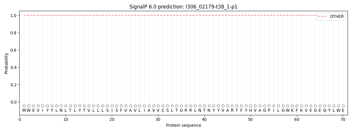
This protein is predicted as OTHER

| Other | SP_Sec_SPI | CS Position |
|---|---|---|
| 1.000077 | 0.000000 |

