You are browsing environment: FUNGIDB
CAZyme Information: HMPREF1541_02589-t46_1-p1
You are here: Home > Sequence: HMPREF1541_02589-t46_1-p1
Basic Information |
Genomic context |
Full Sequence |
Enzyme annotations |
CAZy signature domains |
CDD domains |
CAZyme hits |
PDB hits |
Swiss-Prot hits |
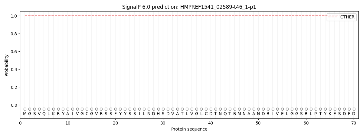
SignalP and Lipop annotations |
TMHMM annotations
Basic Information help
| Species | Cyphellophora europaea | |||||||||||
|---|---|---|---|---|---|---|---|---|---|---|---|---|
| Lineage | Ascomycota; Eurotiomycetes; ; Cyphellophoraceae; Cyphellophora; Cyphellophora europaea | |||||||||||
| CAZyme ID | HMPREF1541_02589-t46_1-p1 | |||||||||||
| CAZy Family | CE1 | |||||||||||
| CAZyme Description | hypothetical protein | |||||||||||
| CAZyme Property |
|
|||||||||||
| Genome Property |
|
|||||||||||
| Gene Location | ||||||||||||
CAZyme Signature Domains help
| Family | Start | End | Evalue | family coverage |
|---|---|---|---|---|
| GH109 | 8 | 171 | 6.3e-25 | 0.40852130325814534 |
CDD Domains download full data without filtering help
| Cdd ID | Domain | E-Value | qStart | qEnd | sStart | sEnd | Domain Description |
|---|---|---|---|---|---|---|---|
| 223745 | MviM | 3.16e-40 | 8 | 223 | 5 | 213 | Predicted dehydrogenase [General function prediction only]. |
| 396129 | GFO_IDH_MocA | 6.68e-17 | 7 | 135 | 1 | 120 | Oxidoreductase family, NAD-binding Rossmann fold. This family of enzymes utilize NADP or NAD. This family is called the GFO/IDH/MOCA family in swiss-prot. |
| 397161 | GFO_IDH_MocA_C | 3.23e-12 | 147 | 435 | 1 | 204 | Oxidoreductase family, C-terminal alpha/beta domain. This family of enzymes utilize NADP or NAD. This family is called the GFO/IDH/MOCA family in swiss-prot. |
| 182305 | PRK10206 | 3.79e-05 | 67 | 165 | 54 | 154 | putative oxidoreductase; Provisional |
| 281446 | NAD_binding_3 | 6.45e-05 | 13 | 133 | 1 | 115 | Homoserine dehydrogenase, NAD binding domain. This domain adopts a Rossmann NAD binding fold. The C-terminal domain of homoserine dehydrogenase contributes a single helix to this structural domain, which is not included in the Pfam model. |
CAZyme Hits help
| Hit ID | E-Value | Query Start | Query End | Hit Start | Hit End |
|---|---|---|---|---|---|
| 8.77e-25 | 8 | 212 | 396 | 604 | |
| 2.91e-13 | 8 | 285 | 5 | 268 | |
| 8.26e-10 | 3 | 392 | 51 | 425 | |
| 1.03e-09 | 4 | 158 | 52 | 206 | |
| 1.03e-09 | 4 | 392 | 52 | 397 |
PDB Hits download full data without filtering help
| Hit ID | E-Value | Query Start | Query End | Hit Start | Hit End | Description |
|---|---|---|---|---|---|---|
| 3.08e-14 | 4 | 217 | 1 | 206 | CRYSTAL STRUCTURE OF NAD-BINDING PROTEIN FROM Listeria innocua [Listeria innocua],3E18_B CRYSTAL STRUCTURE OF NAD-BINDING PROTEIN FROM Listeria innocua [Listeria innocua] |
|
| 5.04e-12 | 8 | 201 | 4 | 186 | The crystal structure of the putative dehydrogenase from Bordetella bronchiseptica RB50 [Bordetella bronchiseptica] |
|
| 2.09e-11 | 8 | 212 | 5 | 191 | Crystal structure of a putative myo-inositol dehydrogenase from Sinorhizobium meliloti 1021 (Target PSI-012312) [Sinorhizobium meliloti 1021],4HKT_B Crystal structure of a putative myo-inositol dehydrogenase from Sinorhizobium meliloti 1021 (Target PSI-012312) [Sinorhizobium meliloti 1021],4HKT_C Crystal structure of a putative myo-inositol dehydrogenase from Sinorhizobium meliloti 1021 (Target PSI-012312) [Sinorhizobium meliloti 1021],4HKT_D Crystal structure of a putative myo-inositol dehydrogenase from Sinorhizobium meliloti 1021 (Target PSI-012312) [Sinorhizobium meliloti 1021] |
|
| 2.43e-10 | 8 | 213 | 15 | 212 | Crystal structure of the WlbA dehydrognase from Chromobactrium violaceum in complex with NADH and UDP-GlcNAcA at 1.50 A resolution [Chromobacterium violaceum] |
|
| 8.25e-10 | 8 | 238 | 32 | 251 | Crystal structure of the WlbA dehydrogenase from Bordetella pertussis in complex with NADH and UDP-GlcNAcA [Bordetella pertussis Tohama I],3Q2K_B Crystal structure of the WlbA dehydrogenase from Bordetella pertussis in complex with NADH and UDP-GlcNAcA [Bordetella pertussis Tohama I],3Q2K_C Crystal structure of the WlbA dehydrogenase from Bordetella pertussis in complex with NADH and UDP-GlcNAcA [Bordetella pertussis Tohama I],3Q2K_D Crystal structure of the WlbA dehydrogenase from Bordetella pertussis in complex with NADH and UDP-GlcNAcA [Bordetella pertussis Tohama I],3Q2K_E Crystal structure of the WlbA dehydrogenase from Bordetella pertussis in complex with NADH and UDP-GlcNAcA [Bordetella pertussis Tohama I],3Q2K_F Crystal structure of the WlbA dehydrogenase from Bordetella pertussis in complex with NADH and UDP-GlcNAcA [Bordetella pertussis Tohama I],3Q2K_G Crystal structure of the WlbA dehydrogenase from Bordetella pertussis in complex with NADH and UDP-GlcNAcA [Bordetella pertussis Tohama I],3Q2K_H Crystal structure of the WlbA dehydrogenase from Bordetella pertussis in complex with NADH and UDP-GlcNAcA [Bordetella pertussis Tohama I],3Q2K_I Crystal structure of the WlbA dehydrogenase from Bordetella pertussis in complex with NADH and UDP-GlcNAcA [Bordetella pertussis Tohama I],3Q2K_J Crystal structure of the WlbA dehydrogenase from Bordetella pertussis in complex with NADH and UDP-GlcNAcA [Bordetella pertussis Tohama I],3Q2K_K Crystal structure of the WlbA dehydrogenase from Bordetella pertussis in complex with NADH and UDP-GlcNAcA [Bordetella pertussis Tohama I],3Q2K_L Crystal structure of the WlbA dehydrogenase from Bordetella pertussis in complex with NADH and UDP-GlcNAcA [Bordetella pertussis Tohama I],3Q2K_M Crystal structure of the WlbA dehydrogenase from Bordetella pertussis in complex with NADH and UDP-GlcNAcA [Bordetella pertussis Tohama I],3Q2K_N Crystal structure of the WlbA dehydrogenase from Bordetella pertussis in complex with NADH and UDP-GlcNAcA [Bordetella pertussis Tohama I],3Q2K_O Crystal structure of the WlbA dehydrogenase from Bordetella pertussis in complex with NADH and UDP-GlcNAcA [Bordetella pertussis Tohama I],3Q2K_P Crystal structure of the WlbA dehydrogenase from Bordetella pertussis in complex with NADH and UDP-GlcNAcA [Bordetella pertussis Tohama I] |
Swiss-Prot Hits download full data without filtering help
| Hit ID | E-Value | Query Start | Query End | Hit Start | Hit End | Description |
|---|---|---|---|---|---|---|
| 5.31e-73 | 11 | 438 | 4 | 422 | Putative oxidoreductase YteT OS=Bacillus subtilis (strain 168) OX=224308 GN=yteT PE=2 SV=1 |
|
| 6.69e-12 | 8 | 158 | 11 | 153 | D-glucoside 3-dehydrogenase OS=Escherichia coli (strain K12) OX=83333 GN=ycjS PE=1 SV=1 |
|
| 4.46e-11 | 8 | 212 | 4 | 190 | Inositol 2-dehydrogenase OS=Rhizobium meliloti (strain 1021) OX=266834 GN=idhA PE=1 SV=2 |
|
| 1.84e-10 | 4 | 392 | 52 | 397 | Glycosyl hydrolase family 109 protein OS=Shewanella baltica (strain OS185) OX=402882 GN=Shew185_2813 PE=3 SV=1 |
|
| 1.84e-10 | 4 | 392 | 52 | 397 | Glycosyl hydrolase family 109 protein OS=Shewanella baltica (strain OS155 / ATCC BAA-1091) OX=325240 GN=Sbal_2793 PE=3 SV=1 |
SignalP and Lipop Annotations help
This protein is predicted as OTHER

| Other | SP_Sec_SPI | CS Position |
|---|---|---|
| 1.000040 | 0.000001 |
