You are browsing environment: FUNGIDB
CAZyme Information: GAQ12176.1
You are here: Home > Sequence: GAQ12176.1
Basic Information |
Genomic context |
Full Sequence |
Enzyme annotations |
CAZy signature domains |
CDD domains |
CAZyme hits |
PDB hits |
Swiss-Prot hits |
SignalP and Lipop annotations |
TMHMM annotations
Basic Information help
| Species | Aspergillus lentulus | |||||||||||
|---|---|---|---|---|---|---|---|---|---|---|---|---|
| Lineage | Ascomycota; Eurotiomycetes; ; Aspergillaceae; Aspergillus; Aspergillus lentulus | |||||||||||
| CAZyme ID | GAQ12176.1 | |||||||||||
| CAZy Family | GT59 | |||||||||||
| CAZyme Description | FAD-binding PCMH-type domain-containing protein [Source:UniProtKB/TrEMBL;Acc:A0A0S7E9V4] | |||||||||||
| CAZyme Property |
|
|||||||||||
| Genome Property |
|
|||||||||||
| Gene Location | ||||||||||||
CAZyme Signature Domains help
| Family | Start | End | Evalue | family coverage |
|---|---|---|---|---|
| AA7 | 390 | 623 | 1.2e-64 | 0.5109170305676856 |
CDD Domains download full data without filtering help
| Cdd ID | Domain | E-Value | qStart | qEnd | sStart | sEnd | Domain Description |
|---|---|---|---|---|---|---|---|
| 187561 | NmrA_like_SDR_a | 3.86e-59 | 28 | 282 | 1 | 234 | NmrA (a transcriptional regulator) and HSCARG (an NADPH sensor) like proteins, atypical (a) SDRs. NmrA and HSCARG like proteins. NmrA is a negative transcriptional regulator of various fungi, involved in the post-translational modulation of the GATA-type transcription factor AreA. NmrA lacks the canonical GXXGXXG NAD-binding motif and has altered residues at the catalytic triad, including a Met instead of the critical Tyr residue. NmrA may bind nucleotides but appears to lack any dehydrogenase activity. HSCARG has been identified as a putative NADP-sensing molecule, and redistributes and restructures in response to NADPH/NADP ratios. Like NmrA, it lacks most of the active site residues of the SDR family, but has an NAD(P)-binding motif similar to the extended SDR family, GXXGXXG. SDRs are a functionally diverse family of oxidoreductases that have a single domain with a structurally conserved Rossmann fold, an NAD(P)(H)-binding region, and a structurally diverse C-terminal region. Sequence identity between different SDR enzymes is typically in the 15-30% range; they catalyze a wide range of activities including the metabolism of steroids, cofactors, carbohydrates, lipids, aromatic compounds, and amino acids, and act in redox sensing. Atypical SDRs are distinct from classical SDRs. Classical SDRs have an TGXXX[AG]XG cofactor binding motif and a YXXXK active site motif, with the Tyr residue of the active site motif serving as a critical catalytic residue (Tyr-151, human 15-hydroxyprostaglandin dehydrogenase numbering). In addition to the Tyr and Lys, there is often an upstream Ser and/or an Asn, contributing to the active site; while substrate binding is in the C-terminal region, which determines specificity. The standard reaction mechanism is a 4-pro-S hydride transfer and proton relay involving the conserved Tyr and Lys, a water molecule stabilized by Asn, and nicotinamide. In addition to the Rossmann fold core region typical of all SDRs, extended SDRs have a less conserved C-terminal extension of approximately 100 amino acids, and typically have a TGXXGXXG cofactor binding motif. Complex (multidomain) SDRs such as ketoreductase domains of fatty acid synthase have a GGXGXXG NAD(P)-binding motif and an altered active site motif (YXXXN). Fungal type ketoacyl reductases have a TGXXXGX(1-2)G NAD(P)-binding motif. |
| 398829 | NmrA | 1.67e-35 | 28 | 278 | 1 | 236 | NmrA-like family. NmrA is a negative transcriptional regulator involved in the post-translational modification of the transcription factor AreA. NmrA is part of a system controlling nitrogen metabolite repression in fungi. This family only contains a few sequences as iteration results in significant matches to other Rossmann fold families. |
| 223354 | GlcD | 1.35e-26 | 396 | 786 | 30 | 451 | FAD/FMN-containing dehydrogenase [Energy production and conversion]. |
| 396238 | FAD_binding_4 | 2.09e-26 | 398 | 533 | 1 | 139 | FAD binding domain. This family consists of various enzymes that use FAD as a co-factor, most of the enzymes are similar to oxygen oxidoreductase. One of the enzymes Vanillyl-alcohol oxidase (VAO) has a solved structure, the alignment includes the FAD binding site, called the PP-loop, between residues 99-110. The FAD molecule is covalently bound in the known structure, however the residue that links to the FAD is not in the alignment. VAO catalyzes the oxidation of a wide variety of substrates, ranging form aromatic amines to 4-alkylphenols. Other members of this family include D-lactate dehydrogenase, this enzyme catalyzes the conversion of D-lactate to pyruvate using FAD as a co-factor; mitomycin radical oxidase, this enzyme oxidizes the reduced form of mitomycins and is involved in mitomycin resistance. This family includes MurB an UDP-N-acetylenolpyruvoylglucosamine reductase enzyme EC:1.1.1.158. This enzyme is involved in the biosynthesis of peptidoglycan. |
| 187651 | NmrA_TMR_like_SDR_a | 2.39e-24 | 28 | 279 | 1 | 224 | NmrA (a transcriptional regulator), HSCARG (an NADPH sensor), and triphenylmethane reductase (TMR) like proteins, atypical (a) SDRs. Atypical SDRs belonging to this subgroup include NmrA, HSCARG, and TMR, these proteins bind NAD(P) but they lack the usual catalytic residues of the SDRs. Atypical SDRs are distinct from classical SDRs. NmrA is a negative transcriptional regulator of various fungi, involved in the post-translational modulation of the GATA-type transcription factor AreA. NmrA lacks the canonical GXXGXXG NAD-binding motif and has altered residues at the catalytic triad, including a Met instead of the critical Tyr residue. NmrA may bind nucleotides but appears to lack any dehydrogenase activity. HSCARG has been identified as a putative NADP-sensing molecule, and redistributes and restructures in response to NADPH/NADP ratios. Like NmrA, it lacks most of the active site residues of the SDR family, but has an NAD(P)-binding motif similar to the extended SDR family, GXXGXXG. TMR, an NADP-binding protein, lacks the active site residues of the SDRs but has a glycine rich NAD(P)-binding motif that matches the extended SDRs. Atypical SDRs include biliverdin IX beta reductase (BVR-B,aka flavin reductase), NMRa (a negative transcriptional regulator of various fungi), progesterone 5-beta-reductase like proteins, phenylcoumaran benzylic ether and pinoresinol-lariciresinol reductases, phenylpropene synthases, eugenol synthase, triphenylmethane reductase, isoflavone reductases, and others. SDRs are a functionally diverse family of oxidoreductases that have a single domain with a structurally conserved Rossmann fold, an NAD(P)(H)-binding region, and a structurally diverse C-terminal region. Sequence identity between different SDR enzymes is typically in the 15-30% range; they catalyze a wide range of activities including the metabolism of steroids, cofactors, carbohydrates, lipids, aromatic compounds, and amino acids, and act in redox sensing. Classical SDRs have an TGXXX[AG]XG cofactor binding motif and a YXXXK active site motif, with the Tyr residue of the active site motif serving as a critical catalytic residue (Tyr-151, human 15-hydroxyprostaglandin dehydrogenase numbering). In addition to the Tyr and Lys, there is often an upstream Ser and/or an Asn, contributing to the active site; while substrate binding is in the C-terminal region, which determines specificity. The standard reaction mechanism is a 4-pro-S hydride transfer and proton relay involving the conserved Tyr and Lys, a water molecule stabilized by Asn, and nicotinamide. In addition to the Rossmann fold core region typical of all SDRs, extended SDRs have a less conserved C-terminal extension of approximately 100 amino acids, and typically have a TGXXGXXG cofactor binding motif. Complex (multidomain) SDRs such as ketoreductase domains of fatty acid synthase have a GGXGXXG NAD(P)-binding motif and an altered active site motif (YXXXN). Fungal type ketoacyl reductases have a TGXXXGX(1-2)G NAD(P)-binding motif. |
CAZyme Hits help
| Hit ID | E-Value | Query Start | Query End | Hit Start | Hit End |
|---|---|---|---|---|---|
| 9.44e-25 | 398 | 568 | 62 | 234 | |
| 2.37e-24 | 398 | 568 | 37 | 209 | |
| 5.88e-23 | 370 | 785 | 35 | 483 | |
| 5.35e-21 | 378 | 568 | 43 | 236 | |
| 6.92e-21 | 379 | 595 | 44 | 266 |
PDB Hits download full data without filtering help
| Hit ID | E-Value | Query Start | Query End | Hit Start | Hit End | Description |
|---|---|---|---|---|---|---|
| 2.96e-44 | 360 | 794 | 1 | 457 | Crystal structure of 6-hydoxy-D-nicotine oxidase from Arthrobacter nicotinovorans. Crystal Form 3 (P1) [Paenarthrobacter nicotinovorans],2BVF_B Crystal structure of 6-hydoxy-D-nicotine oxidase from Arthrobacter nicotinovorans. Crystal Form 3 (P1) [Paenarthrobacter nicotinovorans],2BVG_A Crystal structure of 6-hydoxy-D-nicotine oxidase from Arthrobacter nicotinovorans. Crystal Form 1 (P21) [Paenarthrobacter nicotinovorans],2BVG_B Crystal structure of 6-hydoxy-D-nicotine oxidase from Arthrobacter nicotinovorans. Crystal Form 1 (P21) [Paenarthrobacter nicotinovorans],2BVG_C Crystal structure of 6-hydoxy-D-nicotine oxidase from Arthrobacter nicotinovorans. Crystal Form 1 (P21) [Paenarthrobacter nicotinovorans],2BVG_D Crystal structure of 6-hydoxy-D-nicotine oxidase from Arthrobacter nicotinovorans. Crystal Form 1 (P21) [Paenarthrobacter nicotinovorans],2BVH_A Crystal structure of 6-hydoxy-D-nicotine oxidase from Arthrobacter nicotinovorans. Crystal Form 2 (P21) [Paenarthrobacter nicotinovorans],2BVH_B Crystal structure of 6-hydoxy-D-nicotine oxidase from Arthrobacter nicotinovorans. Crystal Form 2 (P21) [Paenarthrobacter nicotinovorans],2BVH_C Crystal structure of 6-hydoxy-D-nicotine oxidase from Arthrobacter nicotinovorans. Crystal Form 2 (P21) [Paenarthrobacter nicotinovorans],2BVH_D Crystal structure of 6-hydoxy-D-nicotine oxidase from Arthrobacter nicotinovorans. Crystal Form 2 (P21) [Paenarthrobacter nicotinovorans] |
|
| 1.87e-37 | 375 | 785 | 22 | 453 | The crystal structure of EncM V135T mutant [Streptomyces maritimus],6FYG_B The crystal structure of EncM V135T mutant [Streptomyces maritimus],6FYG_C The crystal structure of EncM V135T mutant [Streptomyces maritimus],6FYG_D The crystal structure of EncM V135T mutant [Streptomyces maritimus] |
|
| 2.53e-37 | 375 | 785 | 22 | 453 | The crystal structure of EncM T139V mutant [Streptomyces maritimus],6FYD_B The crystal structure of EncM T139V mutant [Streptomyces maritimus],6FYD_C The crystal structure of EncM T139V mutant [Streptomyces maritimus],6FYD_D The crystal structure of EncM T139V mutant [Streptomyces maritimus] |
|
| 8.45e-37 | 375 | 785 | 22 | 453 | Crystal Structure of EncM (crystallized with 4 mM NADPH) [Streptomyces maritimus],4XLO_B Crystal Structure of EncM (crystallized with 4 mM NADPH) [Streptomyces maritimus],4XLO_C Crystal Structure of EncM (crystallized with 4 mM NADPH) [Streptomyces maritimus],4XLO_D Crystal Structure of EncM (crystallized with 4 mM NADPH) [Streptomyces maritimus],6FOQ_A The crystal structure of EncM complexed with dioxygen under 15 bar of oxygen pressure. [Streptomyces maritimus],6FOQ_B The crystal structure of EncM complexed with dioxygen under 15 bar of oxygen pressure. [Streptomyces maritimus],6FOQ_C The crystal structure of EncM complexed with dioxygen under 15 bar of oxygen pressure. [Streptomyces maritimus],6FOQ_D The crystal structure of EncM complexed with dioxygen under 15 bar of oxygen pressure. [Streptomyces maritimus],6FOW_A The crystal structure of EncM complexed with dioxygen under 10 bar of oxygen pressure. [Streptomyces maritimus],6FOW_B The crystal structure of EncM complexed with dioxygen under 10 bar of oxygen pressure. [Streptomyces maritimus],6FOW_C The crystal structure of EncM complexed with dioxygen under 10 bar of oxygen pressure. [Streptomyces maritimus],6FOW_D The crystal structure of EncM complexed with dioxygen under 10 bar of oxygen pressure. [Streptomyces maritimus],6FP3_A The crystal structure of EncM complexed with dioxygen under 5 bar of oxygen pressure. [Streptomyces maritimus],6FP3_B The crystal structure of EncM complexed with dioxygen under 5 bar of oxygen pressure. [Streptomyces maritimus],6FP3_C The crystal structure of EncM complexed with dioxygen under 5 bar of oxygen pressure. [Streptomyces maritimus],6FP3_D The crystal structure of EncM complexed with dioxygen under 5 bar of oxygen pressure. [Streptomyces maritimus],6FY8_A The crystal structure of EncM bromide soak [Streptomyces maritimus],6FY9_A The crystal structure of EncM complex with xenon under 15 bars Xe pressure [Streptomyces maritimus],6FYA_A The crystal structure of EncM under anaerobic conditions [Streptomyces maritimus],6FYA_B The crystal structure of EncM under anaerobic conditions [Streptomyces maritimus] |
|
| 9.01e-37 | 375 | 785 | 26 | 457 | The crystal structure of EncM [Streptomyces maritimus],3W8W_B The crystal structure of EncM [Streptomyces maritimus],3W8X_A The complex structure of EncM with trifluorotriketide [Streptomyces maritimus],3W8X_B The complex structure of EncM with trifluorotriketide [Streptomyces maritimus],3W8Z_A The complex structure of EncM with hydroxytetraketide [Streptomyces maritimus],3W8Z_B The complex structure of EncM with hydroxytetraketide [Streptomyces maritimus] |
Swiss-Prot Hits download full data without filtering help
| Hit ID | E-Value | Query Start | Query End | Hit Start | Hit End | Description |
|---|---|---|---|---|---|---|
| 5.66e-79 | 21 | 342 | 1 | 323 | NmrA-like family domain-containing oxidoreductase hkm9 OS=Aspergillus hancockii OX=1873369 PE=1 SV=1 |
|
| 3.30e-74 | 21 | 334 | 1 | 315 | NmrA-like family domain-containing oxidoreductase lnaB OS=Aspergillus flavus (strain ATCC 200026 / FGSC A1120 / IAM 13836 / NRRL 3357 / JCM 12722 / SRRC 167) OX=332952 GN=lnaB PE=2 SV=1 |
|
| 1.79e-73 | 21 | 330 | 1 | 312 | NmrA-like family domain-containing oxidoreductase ptmS OS=Penicillium simplicissimum OX=69488 GN=ptmS PE=3 SV=1 |
|
| 2.37e-69 | 28 | 329 | 4 | 303 | NmrA-like family domain-containing oxidoreductase himF OS=Aspergillus japonicus OX=34381 GN=himF PE=3 SV=1 |
|
| 6.60e-64 | 365 | 787 | 4 | 445 | FAD-linked oxidoreductase pyvE OS=Aspergillus violaceofuscus (strain CBS 115571) OX=1450538 GN=pyvE PE=3 SV=1 |
SignalP and Lipop Annotations help
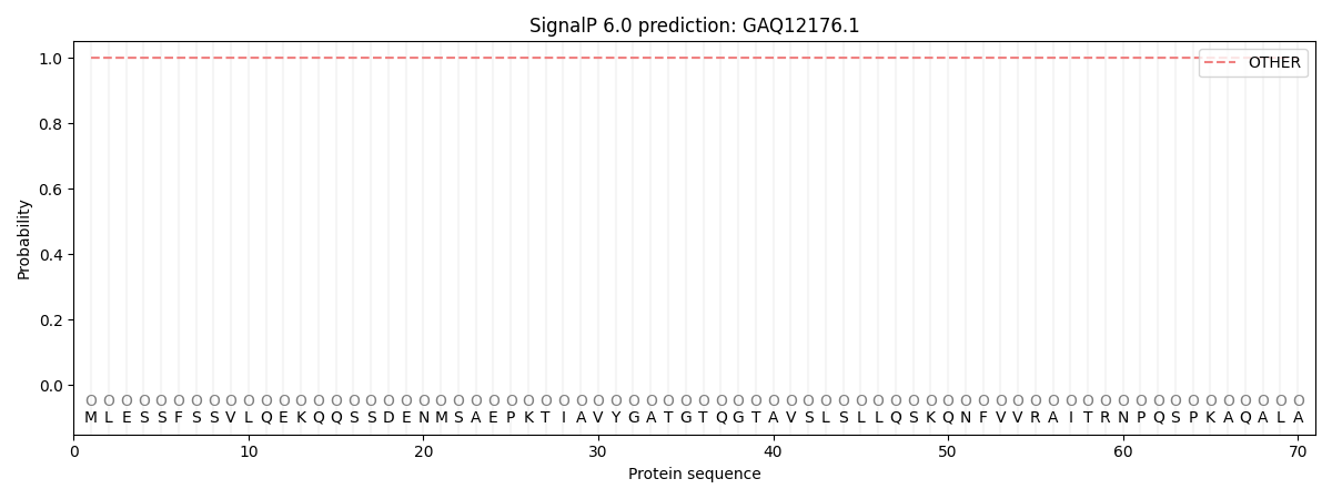
This protein is predicted as OTHER

| Other | SP_Sec_SPI | CS Position |
|---|---|---|
| 1.000072 | 0.000000 |
