You are browsing environment: FUNGIDB
CAZyme Information: GAQ08539.1
You are here: Home > Sequence: GAQ08539.1
Basic Information |
Genomic context |
Full Sequence |
Enzyme annotations |
CAZy signature domains |
CDD domains |
CAZyme hits |
PDB hits |
Swiss-Prot hits |
SignalP and Lipop annotations |
TMHMM annotations
Basic Information help
| Species | Aspergillus lentulus | |||||||||||
|---|---|---|---|---|---|---|---|---|---|---|---|---|
| Lineage | Ascomycota; Eurotiomycetes; ; Aspergillaceae; Aspergillus; Aspergillus lentulus | |||||||||||
| CAZyme ID | GAQ08539.1 | |||||||||||
| CAZy Family | GH38 | |||||||||||
| CAZyme Description | NPCBM domain-containing protein [Source:UniProtKB/TrEMBL;Acc:A0A0S7E1E9] | |||||||||||
| CAZyme Property |
|
|||||||||||
| Genome Property |
|
|||||||||||
| Gene Location | ||||||||||||
Enzyme Prediction help
| EC | 3.2.1.17:4 |
|---|
CAZyme Signature Domains help
| Family | Start | End | Evalue | family coverage |
|---|---|---|---|---|
| GH25 | 228 | 386 | 1.6e-36 | 0.8192090395480226 |
CDD Domains download full data without filtering help
| Cdd ID | Domain | E-Value | qStart | qEnd | sStart | sEnd | Domain Description |
|---|---|---|---|---|---|---|---|
| 119374 | GH25_CH-type | 4.87e-86 | 219 | 404 | 1 | 189 | CH-type (Chalaropsis-type) lysozymes represent one of four functionally-defined classes of peptidoglycan hydrolases (also referred to as endo-N-acetylmuramidases) that cleave bacterial cell wall peptidoglycans. CH-type lysozymes exhibit both lysozyme (acetylmuramidase) and diacetylmuramidase activity. The first member of this family to be described was a muramidase from the fungus Chalaropsis. However, a majority of the CH-type lysozymes are found in bacteriophages and Gram-positive bacteria such as Streptomyces and Clostridium. CH-type lysozymes have a single glycosyl hydrolase family 25 (GH25) domain with an unusual beta/alpha-barrel fold in which the last strand of the barrel is antiparallel to strands beta7 and beta1. Most CH-type lysozymes appear to lack the cell wall-binding domain found in other GH25 muramidases. |
| 395941 | Glyco_hydro_25 | 2.58e-37 | 231 | 380 | 10 | 153 | Glycosyl hydrolases family 25. |
| 119373 | GH25_muramidase | 3.14e-34 | 221 | 379 | 2 | 149 | Endo-N-acetylmuramidases (muramidases) are lysozymes (also referred to as peptidoglycan hydrolases) that degrade bacterial cell walls by catalyzing the hydrolysis of 1,4-beta-linkages between N-acetylmuramic acid and N-acetyl-D-glucosamine residues. This family of muramidases contains a glycosyl hydrolase family 25 (GH25) catalytic domain and is found in bacteria, fungi, slime molds, round worms, protozoans and bacteriophages. The bacteriophage members are referred to as endolysins which are involved in lysing the host cell at the end of the replication cycle to allow release of mature phage particles. Endolysins are typically modular enzymes consisting of a catalytically active domain that hydrolyzes the peptidoglycan cell wall and a cell wall-binding domain that anchors the protein to the cell wall. Endolysins generally have narrow substrate specificities with either intra-species or intra-genus bacteriolytic activity. |
| 187652 | 5beta-POR_like_SDR_a | 2.74e-33 | 458 | 675 | 13 | 254 | progesterone 5-beta-reductase-like proteins (5beta-POR), atypical (a) SDRs. 5beta-POR catalyzes the reduction of progesterone to 5beta-pregnane-3,20-dione in Digitalis plants. This subgroup of atypical-extended SDRs, shares the structure of an extended SDR, but has a different glycine-rich nucleotide binding motif (GXXGXXG) and lacks the YXXXK active site motif of classical and extended SDRs. Tyr-179 and Lys 147 are present in the active site, but not in the usual SDR configuration. Given these differences, it has been proposed that this subfamily represents a new SDR class. Other atypical SDRs include biliverdin IX beta reductase (BVR-B,aka flavin reductase), NMRa (a negative transcriptional regulator of various fungi), phenylcoumaran benzylic ether and pinoresinol-lariciresinol reductases, phenylpropene synthases, eugenol synthase, triphenylmethane reductase, isoflavone reductases, and others. SDRs are a functionally diverse family of oxidoreductases that have a single domain with a structurally conserved Rossmann fold, an NAD(P)(H)-binding region, and a structurally diverse C-terminal region. Sequence identity between different SDR enzymes is typically in the 15-30% range; they catalyze a wide range of activities including the metabolism of steroids, cofactors, carbohydrates, lipids, aromatic compounds, and amino acids, and act in redox sensing. Classical SDRs have an TGXXX[AG]XG cofactor binding motif and a YXXXK active site motif, with the Tyr residue of the active site motif serving as a critical catalytic residue (Tyr-151, human 15-hydroxyprostaglandin dehydrogenase numbering). In addition to the Tyr and Lys, there is often an upstream Ser and/or an Asn, contributing to the active site; while substrate binding is in the C-terminal region, which determines specificity. The standard reaction mechanism is a 4-pro-S hydride transfer and proton relay involving the conserved Tyr and Lys, a water molecule stabilized by Asn, and nicotinamide. In addition to the Rossmann fold core region typical of all SDRs, extended SDRs have a less conserved C-terminal extension of approximately 100 amino acids, and typically have a TGXXGXXG cofactor binding motif. Complex (multidomain) SDRs such as ketoreductase domains of fatty acid synthase have a GGXGXXG NAD(P)-binding motif and an altered active site motif (YXXXN). Fungal type ketoacyl reductases have a TGXXXGX(1-2)G NAD(P)-binding motif. |
| 119384 | GH25_YegX-like | 1.03e-20 | 221 | 390 | 2 | 165 | YegX is an uncharacterized bacterial protein with a glycosyl hydrolase family 25 (GH25) catalytic domain that is similar in sequence to the CH-type (Chalaropsis-type) lysozymes of the GH25 family of endolysins. |
CAZyme Hits help
| Hit ID | E-Value | Query Start | Query End | Hit Start | Hit End |
|---|---|---|---|---|---|
| 6.71e-75 | 217 | 384 | 26 | 195 | |
| 4.42e-70 | 219 | 384 | 31 | 202 | |
| 9.47e-40 | 217 | 403 | 61 | 250 | |
| 9.99e-40 | 231 | 403 | 77 | 253 | |
| 3.29e-39 | 214 | 388 | 71 | 243 |
PDB Hits download full data without filtering help
| Hit ID | E-Value | Query Start | Query End | Hit Start | Hit End | Description |
|---|---|---|---|---|---|---|
| 2.98e-34 | 217 | 399 | 3 | 189 | Crystal structure of the bacterial lysozyme from Streptomyces coelicolor at 1.65 A resolution [Streptomyces coelicolor] |
|
| 3.41e-32 | 221 | 384 | 4 | 166 | Chain A, muramidase [Trichobolus zukalii],6ZMV_B Chain B, muramidase [Trichobolus zukalii] |
|
| 1.47e-30 | 221 | 389 | 4 | 173 | Chain A, muramidase [Sodiomyces alcalophilus] |
|
| 7.69e-28 | 220 | 380 | 5 | 165 | The structure of a family GH25 lysozyme from Aspergillus fumigatus [Aspergillus fumigatus Af293],2X8R_B The structure of a family GH25 lysozyme from Aspergillus fumigatus [Aspergillus fumigatus Af293],2X8R_C The structure of a family GH25 lysozyme from Aspergillus fumigatus [Aspergillus fumigatus Af293],2X8R_D The structure of a family GH25 lysozyme from Aspergillus fumigatus [Aspergillus fumigatus Af293],2X8R_E The structure of a family GH25 lysozyme from Aspergillus fumigatus [Aspergillus fumigatus Af293],2X8R_F The structure of a family GH25 lysozyme from Aspergillus fumigatus [Aspergillus fumigatus Af293] |
Swiss-Prot Hits download full data without filtering help
| Hit ID | E-Value | Query Start | Query End | Hit Start | Hit End | Description |
|---|---|---|---|---|---|---|
| 5.74e-33 | 214 | 399 | 77 | 266 | Lysozyme M1 OS=Streptomyces globisporus OX=1908 GN=acm PE=1 SV=1 |
|
| 3.41e-28 | 220 | 378 | 3 | 164 | N,O-diacetylmuramidase OS=Chalaropsis sp. OX=36534 PE=1 SV=1 |
|
| 2.93e-27 | 220 | 384 | 31 | 194 | N,O-diacetylmuramidase OS=Arthroderma benhamiae (strain ATCC MYA-4681 / CBS 112371) OX=663331 GN=ARB_05911 PE=1 SV=1 |
|
| 4.91e-18 | 457 | 674 | 19 | 285 | Short chain dehydrogenase ausT OS=Aspergillus calidoustus OX=454130 GN=ausT PE=1 SV=1 |
|
| 5.91e-15 | 457 | 670 | 14 | 272 | Short chain dehydrogenase sirQ OS=Leptosphaeria maculans OX=5022 GN=sirQ PE=3 SV=1 |
SignalP and Lipop Annotations help
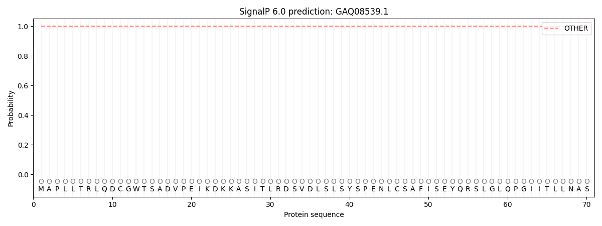
This protein is predicted as OTHER

| Other | SP_Sec_SPI | CS Position |
|---|---|---|
| 1.000056 | 0.000001 |
