You are browsing environment: FUNGIDB
CAZyme Information: EPrPAT00000016604-p1
You are here: Home > Sequence: EPrPAT00000016604-p1
Basic Information |
Genomic context |
Full Sequence |
Enzyme annotations |
CAZy signature domains |
CDD domains |
CAZyme hits |
PDB hits |
Swiss-Prot hits |
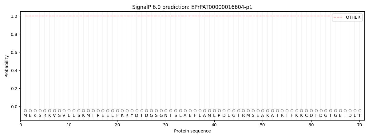
SignalP and Lipop annotations |
TMHMM annotations
Basic Information help
| Species | Pythium aphanidermatum | |||||||||||
|---|---|---|---|---|---|---|---|---|---|---|---|---|
| Lineage | Oomycota; NA; ; Pythiaceae; Pythium; Pythium aphanidermatum | |||||||||||
| CAZyme ID | EPrPAT00000016604-p1 | |||||||||||
| CAZy Family | CE5 | |||||||||||
| CAZyme Description | Regulator of chromosome condensation (RCC1). | |||||||||||
| CAZyme Property |
|
|||||||||||
| Genome Property |
|
|||||||||||
| Gene Location | ||||||||||||
CAZyme Signature Domains help
| Family | Start | End | Evalue | family coverage |
|---|---|---|---|---|
| CBM47 | 1011 | 1149 | 2.9e-19 | 0.9453125 |
CDD Domains download full data without filtering help
| Cdd ID | Domain | E-Value | qStart | qEnd | sStart | sEnd | Domain Description |
|---|---|---|---|---|---|---|---|
| 227511 | ATS1 | 1.32e-22 | 721 | 991 | 192 | 441 | Alpha-tubulin suppressor and related RCC1 domain-containing proteins [Cell cycle control, cell division, chromosome partitioning, Cytoskeleton]. |
| 227511 | ATS1 | 1.71e-20 | 714 | 979 | 54 | 321 | Alpha-tubulin suppressor and related RCC1 domain-containing proteins [Cell cycle control, cell division, chromosome partitioning, Cytoskeleton]. |
| 227455 | FRQ1 | 1.76e-20 | 1 | 159 | 1 | 156 | Ca2+-binding protein, EF-hand superfamily [Signal transduction mechanisms]. |
| 238008 | EFh | 4.33e-16 | 98 | 157 | 3 | 62 | EF-hand, calcium binding motif; A diverse superfamily of calcium sensors and calcium signal modulators; most examples in this alignment model have 2 active canonical EF hands. Ca2+ binding induces a conformational change in the EF-hand motif, leading to the activation or inactivation of target proteins. EF-hands tend to occur in pairs or higher copy numbers. |
| 320060 | EFh_PEF_ALG-2_like | 1.36e-15 | 20 | 164 | 6 | 135 | EF-hand, calcium binding motif, found in homologs of mammalian apoptosis-linked gene 2 protein (ALG-2). The family includes some homologs of mammalian apoptosis-linked gene 2 protein (ALG-2) mainly found in lower eukaryotes, such as a parasitic protist Leishmarua major and a cellular slime mold Dictyostelium discoideum. These homologs contains five EF-hand motifs. Due to the presence of unfavorable residues at the Ca2+-coordinating positions, their non-canonical EF4 and EF5 hands may not bind Ca2+. Two Dictyostelium PEF proteins are the prototypes of this family. They may bind to cytoskeletal proteins and/or signal-transducing proteins localized to detergent-resistant membranes named lipid rafts, and occur as monomers or weak homo- or heterodimers like ALG-2. They can serve as a mediator for Ca2+ signaling-related Dictyostehum programmed cell death (PCD). |
CAZyme Hits help
| Hit ID | E-Value | Query Start | Query End | Hit Start | Hit End |
|---|---|---|---|---|---|
| 1.10e-17 | 998 | 1153 | 131 | 272 | |
| 1.04e-16 | 998 | 1153 | 131 | 272 | |
| 6.29e-16 | 998 | 1155 | 1352 | 1495 | |
| 2.80e-15 | 994 | 1151 | 614 | 761 | |
| 1.36e-13 | 994 | 1153 | 678 | 827 |
PDB Hits download full data without filtering help
| Hit ID | E-Value | Query Start | Query End | Hit Start | Hit End | Description |
|---|---|---|---|---|---|---|
| 3.26e-23 | 726 | 975 | 78 | 298 | Crystal structure of UVB-resistance protein UVR8 [Arabidopsis thaliana],4DNW_B Crystal structure of UVB-resistance protein UVR8 [Arabidopsis thaliana] |
|
| 3.41e-23 | 726 | 975 | 77 | 297 | Crystal structure of UVB photoreceptor UVR8 from Arabidopsis thaliana and UV-induced structural changes at 120K [Arabidopsis thaliana],4NAA_B Crystal structure of UVB photoreceptor UVR8 from Arabidopsis thaliana and UV-induced structural changes at 120K [Arabidopsis thaliana],4NAA_C Crystal structure of UVB photoreceptor UVR8 from Arabidopsis thaliana and UV-induced structural changes at 120K [Arabidopsis thaliana],4NAA_D Crystal structure of UVB photoreceptor UVR8 from Arabidopsis thaliana and UV-induced structural changes at 120K [Arabidopsis thaliana],4NBM_A Crystal structure of UVB photoreceptor UVR8 and light-induced structural changes at 180K [Arabidopsis thaliana],4NBM_B Crystal structure of UVB photoreceptor UVR8 and light-induced structural changes at 180K [Arabidopsis thaliana],4NBM_C Crystal structure of UVB photoreceptor UVR8 and light-induced structural changes at 180K [Arabidopsis thaliana],4NBM_D Crystal structure of UVB photoreceptor UVR8 and light-induced structural changes at 180K [Arabidopsis thaliana],6DD7_A Crystal structure of plant UVB photoreceptor UVR8 from in situ serial Laue diffraction [Arabidopsis thaliana],6DD7_B Crystal structure of plant UVB photoreceptor UVR8 from in situ serial Laue diffraction [Arabidopsis thaliana],6DD7_C Crystal structure of plant UVB photoreceptor UVR8 from in situ serial Laue diffraction [Arabidopsis thaliana],6DD7_D Crystal structure of plant UVB photoreceptor UVR8 from in situ serial Laue diffraction [Arabidopsis thaliana] |
|
| 4.32e-23 | 726 | 975 | 81 | 301 | Chain A, Ultraviolet-B receptor UVR8 [Arabidopsis thaliana],6XZM_B Chain B, Ultraviolet-B receptor UVR8 [Arabidopsis thaliana] |
|
| 5.10e-23 | 726 | 975 | 90 | 310 | Crystal structure of Arabidopsis thaliana UVR8 (UV Resistance locus 8) [Arabidopsis thaliana],4D9S_B Crystal structure of Arabidopsis thaliana UVR8 (UV Resistance locus 8) [Arabidopsis thaliana] |
|
| 5.72e-23 | 726 | 975 | 80 | 300 | Crystal structure of the W285A mutant of UVB-resistance protein UVR8 [Arabidopsis thaliana] |
Swiss-Prot Hits download full data without filtering help
| Hit ID | E-Value | Query Start | Query End | Hit Start | Hit End | Description |
|---|---|---|---|---|---|---|
| 3.96e-22 | 726 | 975 | 89 | 309 | Ultraviolet-B receptor UVR8 OS=Arabidopsis thaliana OX=3702 GN=UVR8 PE=1 SV=1 |
|
| 6.35e-21 | 726 | 980 | 3040 | 3269 | Probable E3 ubiquitin-protein ligase HERC2 OS=Drosophila melanogaster OX=7227 GN=HERC2 PE=1 SV=3 |
|
| 5.84e-20 | 726 | 972 | 43 | 265 | E3 ISG15--protein ligase Herc6 OS=Mus musculus OX=10090 GN=Herc6 PE=2 SV=1 |
|
| 3.61e-19 | 726 | 1055 | 3014 | 3339 | E3 ubiquitin-protein ligase HERC2 OS=Mus musculus OX=10090 GN=Herc2 PE=1 SV=3 |
|
| 6.19e-19 | 726 | 1055 | 3013 | 3338 | E3 ubiquitin-protein ligase HERC2 OS=Homo sapiens OX=9606 GN=HERC2 PE=1 SV=2 |
SignalP and Lipop Annotations help
This protein is predicted as OTHER

| Other | SP_Sec_SPI | CS Position |
|---|---|---|
| 1.000078 | 0.000000 |
