You are browsing environment: FUNGIDB
CAZyme Information: EEU45321.1
You are here: Home > Sequence: EEU45321.1
Basic Information |
Genomic context |
Full Sequence |
Enzyme annotations |
CAZy signature domains |
CDD domains |
CAZyme hits |
PDB hits |
Swiss-Prot hits |
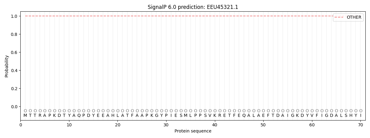
SignalP and Lipop annotations |
TMHMM annotations
Basic Information help
| Species | Fusarium vanettenii | |||||||||||
|---|---|---|---|---|---|---|---|---|---|---|---|---|
| Lineage | Ascomycota; Sordariomycetes; ; Nectriaceae; Fusarium; Fusarium vanettenii | |||||||||||
| CAZyme ID | EEU45321.1 | |||||||||||
| CAZy Family | GT1 | |||||||||||
| CAZyme Description | FAD-binding PCMH-type domain-containing protein [Source:UniProtKB/TrEMBL;Acc:C7YSZ5] | |||||||||||
| CAZyme Property |
|
|||||||||||
| Genome Property |
|
|||||||||||
| Gene Location | ||||||||||||
CAZyme Signature Domains help
| Family | Start | End | Evalue | family coverage |
|---|---|---|---|---|
| AA4 | 34 | 565 | 1e-136 | 0.9865900383141762 |
CDD Domains download full data without filtering help
| Cdd ID | Domain | E-Value | qStart | qEnd | sStart | sEnd | Domain Description |
|---|---|---|---|---|---|---|---|
| 223354 | GlcD | 7.75e-33 | 50 | 556 | 1 | 455 | FAD/FMN-containing dehydrogenase [Energy production and conversion]. |
| 396238 | FAD_binding_4 | 2.83e-28 | 86 | 225 | 1 | 139 | FAD binding domain. This family consists of various enzymes that use FAD as a co-factor, most of the enzymes are similar to oxygen oxidoreductase. One of the enzymes Vanillyl-alcohol oxidase (VAO) has a solved structure, the alignment includes the FAD binding site, called the PP-loop, between residues 99-110. The FAD molecule is covalently bound in the known structure, however the residue that links to the FAD is not in the alignment. VAO catalyzes the oxidation of a wide variety of substrates, ranging form aromatic amines to 4-alkylphenols. Other members of this family include D-lactate dehydrogenase, this enzyme catalyzes the conversion of D-lactate to pyruvate using FAD as a co-factor; mitomycin radical oxidase, this enzyme oxidizes the reduced form of mitomycins and is involved in mitomycin resistance. This family includes MurB an UDP-N-acetylenolpyruvoylglucosamine reductase enzyme EC:1.1.1.158. This enzyme is involved in the biosynthesis of peptidoglycan. |
| 178402 | PLN02805 | 3.58e-11 | 85 | 291 | 133 | 327 | D-lactate dehydrogenase [cytochrome] |
| 183043 | PRK11230 | 5.56e-10 | 80 | 302 | 50 | 261 | glycolate oxidase subunit GlcD; Provisional |
| 273751 | FAD_lactone_ox | 0.004 | 86 | 222 | 15 | 147 | sugar 1,4-lactone oxidases. This model represents a family of at least two different sugar 1,4 lactone oxidases, both involved in synthesizing ascorbic acid or a derivative. These include L-gulonolactone oxidase (EC 1.1.3.8) from rat and D-arabinono-1,4-lactone oxidase (EC 1.1.3.37) from Saccharomyces cerevisiae. Members are proposed to have the cofactor FAD covalently bound at a site specified by Prosite motif PS00862; OX2_COVAL_FAD; 1. |
CAZyme Hits help
| Hit ID | E-Value | Query Start | Query End | Hit Start | Hit End |
|---|---|---|---|---|---|
| 8.03e-293 | 1 | 580 | 1 | 579 | |
| 2.88e-271 | 9 | 568 | 8 | 570 | |
| 9.89e-270 | 1 | 568 | 1 | 567 | |
| 3.86e-252 | 9 | 542 | 8 | 544 | |
| 4.41e-236 | 10 | 565 | 10 | 565 |
PDB Hits download full data without filtering help
| Hit ID | E-Value | Query Start | Query End | Hit Start | Hit End | Description |
|---|---|---|---|---|---|---|
| 2.64e-103 | 35 | 564 | 5 | 522 | Crystal structure of eugenol oxidase in complex with isoeugenol [Rhodococcus jostii RHA1],5FXD_B Crystal structure of eugenol oxidase in complex with isoeugenol [Rhodococcus jostii RHA1],5FXE_A Crystal structure of eugenol oxidase in complex with coniferyl alcohol [Rhodococcus jostii RHA1],5FXE_B Crystal structure of eugenol oxidase in complex with coniferyl alcohol [Rhodococcus jostii RHA1],5FXF_A Crystal structure of eugenol oxidase in complex with benzoate [Rhodococcus jostii RHA1],5FXF_B Crystal structure of eugenol oxidase in complex with benzoate [Rhodococcus jostii RHA1],5FXP_A Crystal structure of eugenol oxidase in complex with vanillin [Rhodococcus jostii RHA1],5FXP_B Crystal structure of eugenol oxidase in complex with vanillin [Rhodococcus jostii RHA1] |
|
| 1.24e-89 | 35 | 565 | 6 | 528 | Chain A, FAD-binding oxidoreductase [Gulosibacter chungangensis],7PBG_B Chain B, FAD-binding oxidoreductase [Gulosibacter chungangensis],7PBI_A Chain A, FAD-binding oxidoreductase [Gulosibacter chungangensis],7PBI_B Chain B, FAD-binding oxidoreductase [Gulosibacter chungangensis],7PBI_C Chain C, FAD-binding oxidoreductase [Gulosibacter chungangensis],7PBI_D Chain D, FAD-binding oxidoreductase [Gulosibacter chungangensis],7PBI_E Chain E, FAD-binding oxidoreductase [Gulosibacter chungangensis],7PBI_F Chain F, FAD-binding oxidoreductase [Gulosibacter chungangensis],7PBI_G Chain G, FAD-binding oxidoreductase [Gulosibacter chungangensis],7PBI_H Chain H, FAD-binding oxidoreductase [Gulosibacter chungangensis] |
|
| 1.18e-87 | 35 | 566 | 12 | 556 | Structure of the D170S/T457E double mutant of vanillyl-alcohol oxidase [Penicillium simplicissimum],1E0Y_B Structure of the D170S/T457E double mutant of vanillyl-alcohol oxidase [Penicillium simplicissimum] |
|
| 3.25e-87 | 35 | 566 | 12 | 556 | Asp170Ser mutant of vanillyl-alcohol oxidase [Penicillium simplicissimum],1DZN_B Asp170Ser mutant of vanillyl-alcohol oxidase [Penicillium simplicissimum] |
|
| 4.56e-87 | 35 | 566 | 12 | 556 | STRUCTURE OF THE OCTAMERIC FLAVOENZYME VANILLYL-ALCOHOL OXIDASE: Ile238Thr Mutant [Penicillium simplicissimum],1W1K_B STRUCTURE OF THE OCTAMERIC FLAVOENZYME VANILLYL-ALCOHOL OXIDASE: Ile238Thr Mutant [Penicillium simplicissimum] |
Swiss-Prot Hits download full data without filtering help
| Hit ID | E-Value | Query Start | Query End | Hit Start | Hit End | Description |
|---|---|---|---|---|---|---|
| 6.48e-86 | 35 | 566 | 12 | 556 | Vanillyl-alcohol oxidase OS=Penicillium simplicissimum OX=69488 GN=VAOA PE=1 SV=1 |
|
| 2.00e-72 | 33 | 558 | 7 | 515 | 4-cresol dehydrogenase [hydroxylating] flavoprotein subunit OS=Pseudomonas putida OX=303 GN=pchF PE=1 SV=3 |
|
| 1.94e-16 | 38 | 300 | 26 | 275 | Probable D-lactate dehydrogenase, mitochondrial OS=Danio rerio OX=7955 GN=ldhd PE=2 SV=1 |
|
| 4.37e-14 | 78 | 300 | 57 | 294 | Probable D-lactate dehydrogenase, mitochondrial OS=Homo sapiens OX=9606 GN=LDHD PE=1 SV=1 |
|
| 4.73e-12 | 85 | 302 | 40 | 247 | Glycolate oxidase subunit GlcD OS=Bacillus subtilis (strain 168) OX=224308 GN=glcD PE=3 SV=1 |
SignalP and Lipop Annotations help
This protein is predicted as OTHER

| Other | SP_Sec_SPI | CS Position |
|---|---|---|
| 1.000053 | 0.000001 |
