You are browsing environment: FUNGIDB
CAZyme Information: CDH51757.1
You are here: Home > Sequence: CDH51757.1
Basic Information |
Genomic context |
Full Sequence |
Enzyme annotations |
CAZy signature domains |
CDD domains |
CAZyme hits |
PDB hits |
Swiss-Prot hits |
SignalP and Lipop annotations |
TMHMM annotations
Basic Information help
| Species | Lichtheimia corymbifera | |||||||||||
|---|---|---|---|---|---|---|---|---|---|---|---|---|
| Lineage | Mucoromycota; Mucoromycetes; ; Lichtheimiaceae; Lichtheimia; Lichtheimia corymbifera | |||||||||||
| CAZyme ID | CDH51757.1 | |||||||||||
| CAZy Family | GH16 | |||||||||||
| CAZyme Description | phosphorylase kinase alphabeta | |||||||||||
| CAZyme Property |
|
|||||||||||
| Genome Property |
|
|||||||||||
| Gene Location | ||||||||||||
CAZyme Signature Domains help
| Family | Start | End | Evalue | family coverage |
|---|---|---|---|---|
| GH15 | 20 | 337 | 8.8e-16 | 0.8088642659279779 |
CDD Domains download full data without filtering help
| Cdd ID | Domain | E-Value | qStart | qEnd | sStart | sEnd | Domain Description |
|---|---|---|---|---|---|---|---|
| 395586 | Glyco_hydro_15 | 8.76e-66 | 17 | 253 | 3 | 226 | Glycosyl hydrolases family 15. In higher organisms this family is represented by phosphorylase kinase subunits. |
| 406202 | ILEI | 5.61e-22 | 1135 | 1235 | 1 | 89 | Interleukin-like EMT inducer. ILEI is a family of proteins found in vertebrates. It is heavily involved in the process of the transition from epithelial to mesenchymal tissue - EMT - during all of embryonic development, cancer progression, metastasis, and chronic inflammation/fibrosis. ILEI is upregulated exclusively at the level of translation, and abnormal ILEI expression, ie cytoplasmic over-expression instead of vesicular localization, is associated with EMT in human cancerous tissue. In order to induce and maintain the EMT of hepatocytes in a TGF-beta-independent fashion ILEI needs the cooperation of oncogenic Ras. |
| 260110 | PANDER_like | 8.83e-20 | 1108 | 1262 | 3 | 149 | Domains similar to the Pancreatic-derived factor. FAM3B or PANDER (PANcreatic DERived factor) has been identifed as a regulator of glucose homeostasis and beta cell function. The protein is expressed in the endocrine pancreas and co-secreted with insulin in response to glucose, particularly under conditions of insulin resistance. The protein had initially been predicted to be a member of the four-helical cytokine family, hence the FAM3B designation. This wider family contains FAM3B and FAM4C, N-terminal domains of N-acetylglucosaminyltransferases, and domains in poorly characterized proteins that have been associated with deafness and the progression of cancer. |
| 260112 | PANDER_like_TMEM2 | 5.40e-18 | 1134 | 1248 | 47 | 149 | PANDER-like domain of the transmembrane protein TMEM2. TMEM2 has been characterized as a transmembrane protein that maps to the DFNB7-DFNB11 deafness locus on human chromosome 9. It contains a domain similar to the Pancreatic-derived factor PANDER, C-terminal to a glycine rich G8-domain. The function of the PANDER-like domain in TMEM2 has not been characterized. |
| 260111 | PANDER_GnT-1_2_like | 2.25e-10 | 1115 | 1245 | 6 | 127 | PANDER-like domain of N-acetylglucosaminyltransferases. O-linked-mannose beta-1,2-N-acetylglucosaminyltransferase 1 participates in O-mannosyl glycosylation and may be responsible for creating GlcNAc(beta1-2)Man(alpha1-)O-Ser/Thr moieties on alpha dystroglycan and other O-mannosylated proteins. The domain characterized by this model lies N-terminal to the catalytic domain. Its function has not been determined. |
CAZyme Hits help
| Hit ID | E-Value | Query Start | Query End | Hit Start | Hit End |
|---|---|---|---|---|---|
| 0.0 | 1 | 1235 | 1 | 1235 | |
| 1.16e-243 | 17 | 1447 | 9 | 1056 | |
| 3.54e-243 | 17 | 1447 | 10 | 1057 | |
| 1.39e-242 | 17 | 1447 | 10 | 1057 | |
| 2.13e-241 | 17 | 1447 | 10 | 1057 |
PDB Hits download full data without filtering help
| Hit ID | E-Value | Query Start | Query End | Hit Start | Hit End | Description |
|---|---|---|---|---|---|---|
| 4.21e-06 | 1115 | 1278 | 13 | 171 | Crystal structure of human protein O-mannose beta-1,2-N-acetylglucosaminyltransferase form II [Homo sapiens],5GGF_B Crystal structure of human protein O-mannose beta-1,2-N-acetylglucosaminyltransferase form II [Homo sapiens],5GGF_C Crystal structure of human protein O-mannose beta-1,2-N-acetylglucosaminyltransferase form II [Homo sapiens],5GGG_A Crystal structure of human protein O-mannose beta-1,2-N-acetylglucosaminyltransferase form I [Homo sapiens],5GGI_A Crystal structure of human protein O-mannose beta-1,2-N-acetylglucosaminyltransferase in complex with Mn, UDP and Mannosyl-peptide [Homo sapiens],5GGI_B Crystal structure of human protein O-mannose beta-1,2-N-acetylglucosaminyltransferase in complex with Mn, UDP and Mannosyl-peptide [Homo sapiens] |
|
| 6.39e-06 | 1115 | 1237 | 18 | 133 | Crystal structure of N-terminal domain of human protein O-mannose beta-1,2-N-acetylglucosaminyltransferase in complex with Man-alpha-pNP [Homo sapiens],5GGJ_B Crystal structure of N-terminal domain of human protein O-mannose beta-1,2-N-acetylglucosaminyltransferase in complex with Man-alpha-pNP [Homo sapiens],5GGK_A Crystal structure of N-terminal domain of human protein O-mannose beta-1,2-N-acetylglucosaminyltransferase in complex with Man-beta-pNP [Homo sapiens],5GGK_B Crystal structure of N-terminal domain of human protein O-mannose beta-1,2-N-acetylglucosaminyltransferase in complex with Man-beta-pNP [Homo sapiens],5GGL_A Crystal structure of N-terminal domain of human protein O-mannose beta-1,2-N-acetylglucosaminyltransferase in complex with GlcNAc-alpha-pNP [Homo sapiens],5GGL_B Crystal structure of N-terminal domain of human protein O-mannose beta-1,2-N-acetylglucosaminyltransferase in complex with GlcNAc-alpha-pNP [Homo sapiens],5GGN_A Crystal structure of N-terminal domain of human protein O-mannose beta-1,2-N-acetylglucosaminyltransferase in complex with GlcNAc-beta-pNP [Homo sapiens],5GGN_B Crystal structure of N-terminal domain of human protein O-mannose beta-1,2-N-acetylglucosaminyltransferase in complex with GlcNAc-beta-pNP [Homo sapiens],5GGO_A Crystal structure of N-terminal domain of human protein O-mannose beta-1,2-N-acetylglucosaminyltransferase in complex with GalNac-beta1,3-GlcNAc-beta-pNP [Homo sapiens],5GGO_B Crystal structure of N-terminal domain of human protein O-mannose beta-1,2-N-acetylglucosaminyltransferase in complex with GalNac-beta1,3-GlcNAc-beta-pNP [Homo sapiens],5GGP_A Crystal structure of N-terminal domain of human protein O-mannose beta-1,2-N-acetylglucosaminyltransferase in complex with GlcNAc-beta1,2-Man-peptide [Homo sapiens],5GGP_B Crystal structure of N-terminal domain of human protein O-mannose beta-1,2-N-acetylglucosaminyltransferase in complex with GlcNAc-beta1,2-Man-peptide [Homo sapiens] |
Swiss-Prot Hits download full data without filtering help
| Hit ID | E-Value | Query Start | Query End | Hit Start | Hit End | Description |
|---|---|---|---|---|---|---|
| 3.66e-220 | 17 | 1448 | 10 | 1225 | Phosphorylase b kinase regulatory subunit alpha, skeletal muscle isoform OS=Mus musculus OX=10090 GN=Phka1 PE=1 SV=3 |
|
| 1.37e-217 | 17 | 1452 | 10 | 1223 | Phosphorylase b kinase regulatory subunit alpha, liver isoform OS=Oryctolagus cuniculus OX=9986 GN=PHKA2 PE=2 SV=1 |
|
| 1.17e-215 | 17 | 1448 | 10 | 1221 | Phosphorylase b kinase regulatory subunit alpha, skeletal muscle isoform OS=Oryctolagus cuniculus OX=9986 GN=PHKA1 PE=1 SV=1 |
|
| 3.05e-215 | 17 | 1452 | 10 | 1223 | Phosphorylase b kinase regulatory subunit alpha, liver isoform OS=Homo sapiens OX=9606 GN=PHKA2 PE=1 SV=1 |
|
| 4.55e-214 | 17 | 1452 | 10 | 1223 | Phosphorylase b kinase regulatory subunit alpha, liver isoform OS=Mus musculus OX=10090 GN=Phka2 PE=1 SV=1 |
SignalP and Lipop Annotations help
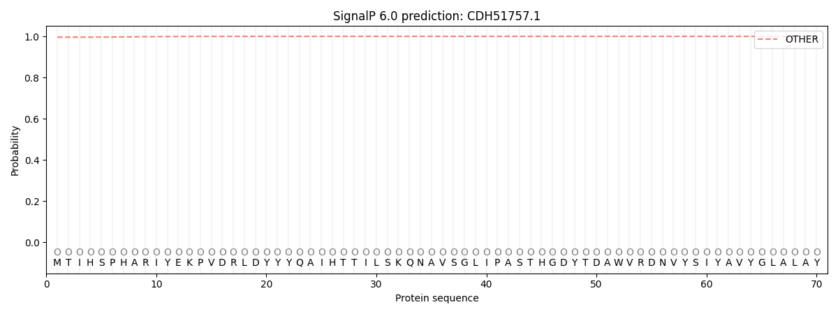
This protein is predicted as OTHER

| Other | SP_Sec_SPI | CS Position |
|---|---|---|
| 0.996724 | 0.003293 |
