You are browsing environment: FUNGIDB
CAZyme Information: ASPACDRAFT_51732-t33_1-p1
You are here: Home > Sequence: ASPACDRAFT_51732-t33_1-p1
Basic Information |
Genomic context |
Full Sequence |
Enzyme annotations |
CAZy signature domains |
CDD domains |
CAZyme hits |
PDB hits |
Swiss-Prot hits |
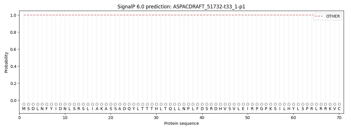
SignalP and Lipop annotations |
TMHMM annotations
Basic Information help
| Species | Aspergillus aculeatus | |||||||||||
|---|---|---|---|---|---|---|---|---|---|---|---|---|
| Lineage | Ascomycota; Eurotiomycetes; ; Aspergillaceae; Aspergillus; Aspergillus aculeatus | |||||||||||
| CAZyme ID | ASPACDRAFT_51732-t33_1-p1 | |||||||||||
| CAZy Family | GH55 | |||||||||||
| CAZyme Description | hypothetical protein | |||||||||||
| CAZyme Property |
|
|||||||||||
| Genome Property |
|
|||||||||||
| Gene Location | ||||||||||||
CAZyme Signature Domains help
| Family | Start | End | Evalue | family coverage |
|---|---|---|---|---|
| AA7 | 282 | 517 | 9e-41 | 0.462882096069869 |
CDD Domains download full data without filtering help
| Cdd ID | Domain | E-Value | qStart | qEnd | sStart | sEnd | Domain Description |
|---|---|---|---|---|---|---|---|
| 235028 | PRK02304 | 2.58e-32 | 926 | 1122 | 1 | 175 | adenine phosphoribosyltransferase; Provisional |
| 398111 | P-mevalo_kinase | 6.26e-23 | 734 | 849 | 1 | 111 | Phosphomevalonate kinase. Phosphomevalonate kinase (EC:2.7.4.2) catalyzes the phosphorylation of 5-phosphomevalonate into 5-diphosphomevalonate, an essential step in isoprenoid biosynthesis via the mevalonate pathway. This family represents the animal type of the enzyme. The other is the ERG8 type, found in plants and fungi, and some bacteria (see pfam00288). |
| 223577 | Apt | 2.23e-22 | 924 | 1122 | 1 | 179 | Adenine/guanine phosphoribosyltransferase or related PRPP-binding protein [Nucleotide transport and metabolism]. |
| 177930 | PLN02293 | 4.92e-20 | 925 | 1104 | 11 | 173 | adenine phosphoribosyltransferase |
| 396238 | FAD_binding_4 | 7.68e-17 | 282 | 413 | 3 | 129 | FAD binding domain. This family consists of various enzymes that use FAD as a co-factor, most of the enzymes are similar to oxygen oxidoreductase. One of the enzymes Vanillyl-alcohol oxidase (VAO) has a solved structure, the alignment includes the FAD binding site, called the PP-loop, between residues 99-110. The FAD molecule is covalently bound in the known structure, however the residue that links to the FAD is not in the alignment. VAO catalyzes the oxidation of a wide variety of substrates, ranging form aromatic amines to 4-alkylphenols. Other members of this family include D-lactate dehydrogenase, this enzyme catalyzes the conversion of D-lactate to pyruvate using FAD as a co-factor; mitomycin radical oxidase, this enzyme oxidizes the reduced form of mitomycins and is involved in mitomycin resistance. This family includes MurB an UDP-N-acetylenolpyruvoylglucosamine reductase enzyme EC:1.1.1.158. This enzyme is involved in the biosynthesis of peptidoglycan. |
CAZyme Hits help
| Hit ID | E-Value | Query Start | Query End | Hit Start | Hit End |
|---|---|---|---|---|---|
| 3.13e-15 | 276 | 715 | 116 | 540 | |
| 2.98e-13 | 275 | 476 | 64 | 243 | |
| 3.06e-13 | 276 | 715 | 72 | 511 | |
| 5.12e-13 | 276 | 510 | 58 | 273 | |
| 6.77e-13 | 276 | 501 | 56 | 262 |
PDB Hits download full data without filtering help
| Hit ID | E-Value | Query Start | Query End | Hit Start | Hit End | Description |
|---|---|---|---|---|---|---|
| 3.97e-17 | 277 | 469 | 36 | 206 | Crystal structure of 6-hydoxy-D-nicotine oxidase from Arthrobacter nicotinovorans. Crystal Form 3 (P1) [Paenarthrobacter nicotinovorans],2BVF_B Crystal structure of 6-hydoxy-D-nicotine oxidase from Arthrobacter nicotinovorans. Crystal Form 3 (P1) [Paenarthrobacter nicotinovorans],2BVG_A Crystal structure of 6-hydoxy-D-nicotine oxidase from Arthrobacter nicotinovorans. Crystal Form 1 (P21) [Paenarthrobacter nicotinovorans],2BVG_B Crystal structure of 6-hydoxy-D-nicotine oxidase from Arthrobacter nicotinovorans. Crystal Form 1 (P21) [Paenarthrobacter nicotinovorans],2BVG_C Crystal structure of 6-hydoxy-D-nicotine oxidase from Arthrobacter nicotinovorans. Crystal Form 1 (P21) [Paenarthrobacter nicotinovorans],2BVG_D Crystal structure of 6-hydoxy-D-nicotine oxidase from Arthrobacter nicotinovorans. Crystal Form 1 (P21) [Paenarthrobacter nicotinovorans],2BVH_A Crystal structure of 6-hydoxy-D-nicotine oxidase from Arthrobacter nicotinovorans. Crystal Form 2 (P21) [Paenarthrobacter nicotinovorans],2BVH_B Crystal structure of 6-hydoxy-D-nicotine oxidase from Arthrobacter nicotinovorans. Crystal Form 2 (P21) [Paenarthrobacter nicotinovorans],2BVH_C Crystal structure of 6-hydoxy-D-nicotine oxidase from Arthrobacter nicotinovorans. Crystal Form 2 (P21) [Paenarthrobacter nicotinovorans],2BVH_D Crystal structure of 6-hydoxy-D-nicotine oxidase from Arthrobacter nicotinovorans. Crystal Form 2 (P21) [Paenarthrobacter nicotinovorans] |
|
| 5.30e-16 | 923 | 1104 | 1 | 168 | Crystal Structure of Mutant R89Q of human Adenine phosphoribosyltransferase [Homo sapiens] |
|
| 5.30e-16 | 923 | 1104 | 1 | 168 | Human Adenine Phosphoribosyltransferase [Homo sapiens],1ZN7_A Human Adenine Phosphoribosyltransferase Complexed with PRPP, ADE and R5P [Homo sapiens],1ZN7_B Human Adenine Phosphoribosyltransferase Complexed with PRPP, ADE and R5P [Homo sapiens],1ZN8_A Human Adenine Phosphoribosyltransferase Complexed with AMP, in Space Group P1 at 1.76 A Resolution [Homo sapiens],1ZN8_B Human Adenine Phosphoribosyltransferase Complexed with AMP, in Space Group P1 at 1.76 A Resolution [Homo sapiens],1ZN9_A Human Adenine Phosphoribosyltransferase in Apo and AMP Complexed Forms [Homo sapiens],1ZN9_B Human Adenine Phosphoribosyltransferase in Apo and AMP Complexed Forms [Homo sapiens] |
|
| 5.30e-16 | 923 | 1104 | 1 | 168 | Crystal Structure of F173G Mutant of Human APRT [Homo sapiens],4X45_B Crystal Structure of F173G Mutant of Human APRT [Homo sapiens] |
|
| 9.35e-16 | 925 | 1104 | 1 | 166 | Crystal Structure of Human APRT wild type in complex with PRPP and Mg2+ [Homo sapiens],6FCH_B Crystal Structure of Human APRT wild type in complex with PRPP and Mg2+ [Homo sapiens],6FCL_A Crystal Structure of Human APRT wild type in complex with AMP [Homo sapiens],6FCL_B Crystal Structure of Human APRT wild type in complex with AMP [Homo sapiens],6HGP_A Crystal Structure of Human APRT wild type in complex with Phosphate ion. [Homo sapiens],6HGP_B Crystal Structure of Human APRT wild type in complex with Phosphate ion. [Homo sapiens],6HGR_A Crystal Structure of Human APRT wild type in complex with IMP [Homo sapiens],6HGR_B Crystal Structure of Human APRT wild type in complex with IMP [Homo sapiens],6HGS_A Crystal Structure of Human APRT wild type in complex with GMP [Homo sapiens],6HGS_B Crystal Structure of Human APRT wild type in complex with GMP [Homo sapiens] |
Swiss-Prot Hits download full data without filtering help
| Hit ID | E-Value | Query Start | Query End | Hit Start | Hit End | Description |
|---|---|---|---|---|---|---|
| 1.20e-27 | 277 | 714 | 45 | 439 | FAD-linked oxidoreductase DDB_G0289697 OS=Dictyostelium discoideum OX=44689 GN=DDB_G0289697 PE=2 SV=1 |
|
| 1.06e-19 | 925 | 1121 | 3 | 180 | Adenine phosphoribosyltransferase OS=Rattus norvegicus OX=10116 GN=Aprt PE=1 SV=1 |
|
| 1.06e-19 | 923 | 1121 | 1 | 180 | Adenine phosphoribosyltransferase OS=Cricetulus griseus OX=10029 GN=APRT PE=3 SV=2 |
|
| 1.96e-19 | 925 | 1121 | 3 | 180 | Adenine phosphoribosyltransferase OS=Mastomys natalensis OX=10112 GN=APRT PE=3 SV=1 |
|
| 6.73e-19 | 925 | 1121 | 3 | 180 | Adenine phosphoribosyltransferase OS=Mus spicilegus OX=10103 GN=Aprt PE=3 SV=1 |
SignalP and Lipop Annotations help
This protein is predicted as OTHER

| Other | SP_Sec_SPI | CS Position |
|---|---|---|
| 1.000056 | 0.000000 |
