You are browsing environment: FUNGIDB
CAZyme Information: APA07661.1
You are here: Home > Sequence: APA07661.1
Basic Information |
Genomic context |
Full Sequence |
Enzyme annotations |
CAZy signature domains |
CDD domains |
CAZyme hits |
PDB hits |
Swiss-Prot hits |
SignalP and Lipop annotations |
TMHMM annotations
Basic Information help
| Species | Sclerotinia sclerotiorum | |||||||||||
|---|---|---|---|---|---|---|---|---|---|---|---|---|
| Lineage | Ascomycota; Leotiomycetes; ; Sclerotiniaceae; Sclerotinia; Sclerotinia sclerotiorum | |||||||||||
| CAZyme ID | APA07661.1 | |||||||||||
| CAZy Family | CE8 | |||||||||||
| CAZyme Description | unspecified product | |||||||||||
| CAZyme Property |
|
|||||||||||
| Genome Property |
|
|||||||||||
| Gene Location | ||||||||||||
CAZyme Signature Domains help
| Family | Start | End | Evalue | family coverage |
|---|---|---|---|---|
| AA7 | 311 | 546 | 1.6e-42 | 0.4585152838427948 |
CDD Domains download full data without filtering help
| Cdd ID | Domain | E-Value | qStart | qEnd | sStart | sEnd | Domain Description |
|---|---|---|---|---|---|---|---|
| 235028 | PRK02304 | 2.38e-37 | 965 | 1157 | 1 | 175 | adenine phosphoribosyltransferase; Provisional |
| 223577 | Apt | 3.34e-26 | 969 | 1157 | 7 | 179 | Adenine/guanine phosphoribosyltransferase or related PRPP-binding protein [Nucleotide transport and metabolism]. |
| 177930 | PLN02293 | 2.26e-21 | 967 | 1139 | 14 | 173 | adenine phosphoribosyltransferase |
| 223354 | GlcD | 1.92e-20 | 295 | 750 | 14 | 450 | FAD/FMN-containing dehydrogenase [Energy production and conversion]. |
| 396238 | FAD_binding_4 | 3.31e-19 | 313 | 456 | 1 | 136 | FAD binding domain. This family consists of various enzymes that use FAD as a co-factor, most of the enzymes are similar to oxygen oxidoreductase. One of the enzymes Vanillyl-alcohol oxidase (VAO) has a solved structure, the alignment includes the FAD binding site, called the PP-loop, between residues 99-110. The FAD molecule is covalently bound in the known structure, however the residue that links to the FAD is not in the alignment. VAO catalyzes the oxidation of a wide variety of substrates, ranging form aromatic amines to 4-alkylphenols. Other members of this family include D-lactate dehydrogenase, this enzyme catalyzes the conversion of D-lactate to pyruvate using FAD as a co-factor; mitomycin radical oxidase, this enzyme oxidizes the reduced form of mitomycins and is involved in mitomycin resistance. This family includes MurB an UDP-N-acetylenolpyruvoylglucosamine reductase enzyme EC:1.1.1.158. This enzyme is involved in the biosynthesis of peptidoglycan. |
CAZyme Hits help
| Hit ID | E-Value | Query Start | Query End | Hit Start | Hit End |
|---|---|---|---|---|---|
| 3.63e-16 | 303 | 752 | 59 | 491 | |
| 4.89e-16 | 303 | 752 | 62 | 493 | |
| 6.26e-16 | 303 | 749 | 52 | 483 | |
| 1.10e-14 | 301 | 504 | 56 | 238 | |
| 1.26e-14 | 306 | 748 | 32 | 462 |
PDB Hits download full data without filtering help
| Hit ID | E-Value | Query Start | Query End | Hit Start | Hit End | Description |
|---|---|---|---|---|---|---|
| 5.77e-20 | 306 | 504 | 31 | 206 | Crystal structure of 6-hydoxy-D-nicotine oxidase from Arthrobacter nicotinovorans. Crystal Form 3 (P1) [Paenarthrobacter nicotinovorans],2BVF_B Crystal structure of 6-hydoxy-D-nicotine oxidase from Arthrobacter nicotinovorans. Crystal Form 3 (P1) [Paenarthrobacter nicotinovorans],2BVG_A Crystal structure of 6-hydoxy-D-nicotine oxidase from Arthrobacter nicotinovorans. Crystal Form 1 (P21) [Paenarthrobacter nicotinovorans],2BVG_B Crystal structure of 6-hydoxy-D-nicotine oxidase from Arthrobacter nicotinovorans. Crystal Form 1 (P21) [Paenarthrobacter nicotinovorans],2BVG_C Crystal structure of 6-hydoxy-D-nicotine oxidase from Arthrobacter nicotinovorans. Crystal Form 1 (P21) [Paenarthrobacter nicotinovorans],2BVG_D Crystal structure of 6-hydoxy-D-nicotine oxidase from Arthrobacter nicotinovorans. Crystal Form 1 (P21) [Paenarthrobacter nicotinovorans],2BVH_A Crystal structure of 6-hydoxy-D-nicotine oxidase from Arthrobacter nicotinovorans. Crystal Form 2 (P21) [Paenarthrobacter nicotinovorans],2BVH_B Crystal structure of 6-hydoxy-D-nicotine oxidase from Arthrobacter nicotinovorans. Crystal Form 2 (P21) [Paenarthrobacter nicotinovorans],2BVH_C Crystal structure of 6-hydoxy-D-nicotine oxidase from Arthrobacter nicotinovorans. Crystal Form 2 (P21) [Paenarthrobacter nicotinovorans],2BVH_D Crystal structure of 6-hydoxy-D-nicotine oxidase from Arthrobacter nicotinovorans. Crystal Form 2 (P21) [Paenarthrobacter nicotinovorans] |
|
| 6.33e-19 | 285 | 762 | 14 | 473 | Crystal structure of carbohydrate oxidase from Microdochium nivale [Microdochium nivale],3RJA_A Crystal structure of carbohydrate oxidase from Microdochium nivale in complex with substrate analogue [Microdochium nivale] |
|
| 4.10e-18 | 967 | 1157 | 6 | 180 | Crystal structure of an APRT from Yersinia pseudotuberculosis in complex with AMP. [Yersinia pseudotuberculosis IP 32953] |
|
| 4.75e-18 | 967 | 1157 | 12 | 186 | Crystal structure of adenine phosphoribosyltransferase from Yersinia pseudotuberculosis. [Yersinia pseudotuberculosis IP 32953],5Y07_A Crystal structure of adenine phosphoribosyltransferase from Yersinia pseudotuberculosis with PRPP. [Yersinia pseudotuberculosis IP 32953],5Y07_B Crystal structure of adenine phosphoribosyltransferase from Yersinia pseudotuberculosis with PRPP. [Yersinia pseudotuberculosis IP 32953],5Y4A_A Cadmium directed assembly of adenine phosphoribosyltransferase from Yersinia pseudotuberculosis. [Yersinia pseudotuberculosis IP 32953],5Y4A_B Cadmium directed assembly of adenine phosphoribosyltransferase from Yersinia pseudotuberculosis. [Yersinia pseudotuberculosis IP 32953],5ZC7_A Crystal structure of APRT from Y. pseudotuberculosis with bound adenine (P63 space group). [Yersinia pseudotuberculosis IP 32953],5ZC7_B Crystal structure of APRT from Y. pseudotuberculosis with bound adenine (P63 space group). [Yersinia pseudotuberculosis IP 32953],5ZMI_A Crystal structure of APRT from Y. pseudotuberculosis in complex with adenine. [Yersinia pseudotuberculosis IP 32953],5ZNQ_A Crystal structure of APRT from Y. pseudotuberculosis with bound adenine (P21 space group). [Yersinia pseudotuberculosis IP 32953],5ZNQ_B Crystal structure of APRT from Y. pseudotuberculosis with bound adenine (P21 space group). [Yersinia pseudotuberculosis IP 32953],5ZOC_A Crystal structure of APRT from Y. pseudotuberculosis with bound adenine (C2 space group). [Yersinia pseudotuberculosis IP 32953] |
|
| 3.22e-17 | 967 | 1157 | 15 | 189 | Crystal structure of project JW0458 from Escherichia coli [Escherichia coli K-12],2DY0_B Crystal structure of project JW0458 from Escherichia coli [Escherichia coli K-12] |
Swiss-Prot Hits download full data without filtering help
| Hit ID | E-Value | Query Start | Query End | Hit Start | Hit End | Description |
|---|---|---|---|---|---|---|
| 1.12e-29 | 304 | 748 | 38 | 439 | FAD-linked oxidoreductase DDB_G0289697 OS=Dictyostelium discoideum OX=44689 GN=DDB_G0289697 PE=2 SV=1 |
|
| 5.81e-22 | 963 | 1143 | 1 | 168 | Adenine phosphoribosyltransferase OS=Actinobacillus succinogenes (strain ATCC 55618 / DSM 22257 / CCUG 43843 / 130Z) OX=339671 GN=apt PE=3 SV=1 |
|
| 2.20e-21 | 970 | 1144 | 5 | 166 | Adenine phosphoribosyltransferase OS=Roseiflexus castenholzii (strain DSM 13941 / HLO8) OX=383372 GN=apt PE=3 SV=1 |
|
| 4.07e-21 | 968 | 1144 | 3 | 166 | Adenine phosphoribosyltransferase OS=Roseiflexus sp. (strain RS-1) OX=357808 GN=apt PE=3 SV=1 |
|
| 1.06e-20 | 970 | 1126 | 13 | 158 | Adenine phosphoribosyltransferase OS=Pectobacterium atrosepticum (strain SCRI 1043 / ATCC BAA-672) OX=218491 GN=apt PE=3 SV=1 |
SignalP and Lipop Annotations help
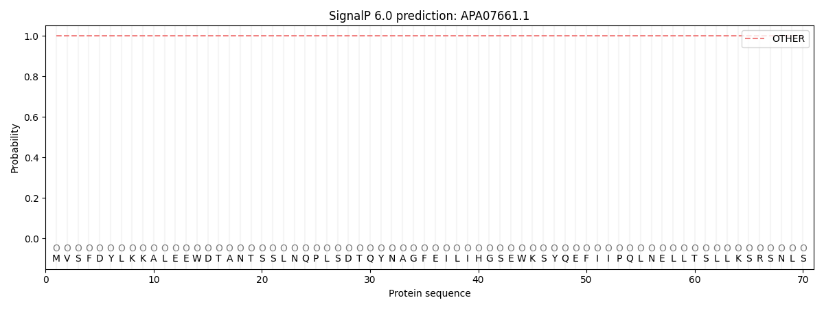
This protein is predicted as OTHER

| Other | SP_Sec_SPI | CS Position |
|---|---|---|
| 1.000050 | 0.000000 |
