You are browsing environment: FUNGIDB
CAZyme Information: AKAW_11278-t41_1-p1
You are here: Home > Sequence: AKAW_11278-t41_1-p1
Basic Information |
Genomic context |
Full Sequence |
Enzyme annotations |
CAZy signature domains |
CDD domains |
CAZyme hits |
PDB hits |
Swiss-Prot hits |
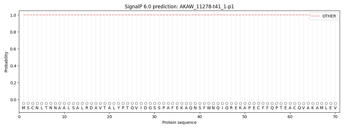
SignalP and Lipop annotations |
TMHMM annotations
Basic Information help
| Species | Aspergillus luchuensis | |||||||||||
|---|---|---|---|---|---|---|---|---|---|---|---|---|
| Lineage | Ascomycota; Eurotiomycetes; ; Aspergillaceae; Aspergillus; Aspergillus luchuensis | |||||||||||
| CAZyme ID | AKAW_11278-t41_1-p1 | |||||||||||
| CAZy Family | GT57 | |||||||||||
| CAZyme Description | FAD binding domain-containing protein | |||||||||||
| CAZyme Property |
|
|||||||||||
| Genome Property |
|
|||||||||||
| Gene Location | ||||||||||||
CAZyme Signature Domains help
| Family | Start | End | Evalue | family coverage |
|---|---|---|---|---|
| AA7 | 41 | 251 | 2.3e-54 | 0.4585152838427948 |
CDD Domains download full data without filtering help
| Cdd ID | Domain | E-Value | qStart | qEnd | sStart | sEnd | Domain Description |
|---|---|---|---|---|---|---|---|
| 396238 | FAD_binding_4 | 2.96e-23 | 51 | 187 | 1 | 139 | FAD binding domain. This family consists of various enzymes that use FAD as a co-factor, most of the enzymes are similar to oxygen oxidoreductase. One of the enzymes Vanillyl-alcohol oxidase (VAO) has a solved structure, the alignment includes the FAD binding site, called the PP-loop, between residues 99-110. The FAD molecule is covalently bound in the known structure, however the residue that links to the FAD is not in the alignment. VAO catalyzes the oxidation of a wide variety of substrates, ranging form aromatic amines to 4-alkylphenols. Other members of this family include D-lactate dehydrogenase, this enzyme catalyzes the conversion of D-lactate to pyruvate using FAD as a co-factor; mitomycin radical oxidase, this enzyme oxidizes the reduced form of mitomycins and is involved in mitomycin resistance. This family includes MurB an UDP-N-acetylenolpyruvoylglucosamine reductase enzyme EC:1.1.1.158. This enzyme is involved in the biosynthesis of peptidoglycan. |
| 223354 | GlcD | 1.87e-20 | 42 | 455 | 23 | 457 | FAD/FMN-containing dehydrogenase [Energy production and conversion]. |
| 100011 | YjgF_YER057c_UK114_like_6 | 3.31e-11 | 442 | 516 | 31 | 119 | This group of proteins belong to a large family of YjgF/YER057c/UK114-like proteins present in bacteria, archaea, and eukaryotes with no definitive function. The conserved domain is similar in structure to chorismate mutase but there is no sequence similarity and no functional connection. Members of this family have been implicated in isoleucine (Yeo7, Ibm1, aldR) and purine (YjgF) biosynthesis, as well as threonine anaerobic degradation (tdcF) and mitochondrial DNA maintenance (Ibm1). This domain homotrimerizes forming a distinct intersubunit cavity that may serve as a small molecule binding site. |
| 223329 | RidA | 5.59e-09 | 443 | 520 | 38 | 130 | Enamine deaminase RidA, house cleaning of reactive enamine intermediates, YjgF/YER057c/UK114 family [Defense mechanisms]. |
| 273751 | FAD_lactone_ox | 1.96e-07 | 51 | 218 | 15 | 180 | sugar 1,4-lactone oxidases. This model represents a family of at least two different sugar 1,4 lactone oxidases, both involved in synthesizing ascorbic acid or a derivative. These include L-gulonolactone oxidase (EC 1.1.3.8) from rat and D-arabinono-1,4-lactone oxidase (EC 1.1.3.37) from Saccharomyces cerevisiae. Members are proposed to have the cofactor FAD covalently bound at a site specified by Prosite motif PS00862; OX2_COVAL_FAD; 1. |
CAZyme Hits help
| Hit ID | E-Value | Query Start | Query End | Hit Start | Hit End |
|---|---|---|---|---|---|
| 2.56e-16 | 51 | 221 | 48 | 219 | |
| 9.72e-16 | 29 | 337 | 44 | 370 | |
| 4.16e-15 | 51 | 218 | 90 | 257 | |
| 4.21e-14 | 62 | 215 | 22 | 176 | |
| 6.21e-14 | 51 | 215 | 63 | 228 |
PDB Hits download full data without filtering help
| Hit ID | E-Value | Query Start | Query End | Hit Start | Hit End | Description |
|---|---|---|---|---|---|---|
| 2.68e-18 | 17 | 217 | 13 | 209 | The crystal structure of EncM T139V mutant [Streptomyces maritimus],6FYD_B The crystal structure of EncM T139V mutant [Streptomyces maritimus],6FYD_C The crystal structure of EncM T139V mutant [Streptomyces maritimus],6FYD_D The crystal structure of EncM T139V mutant [Streptomyces maritimus] |
|
| 4.76e-18 | 17 | 217 | 13 | 209 | The crystal structure of EncM V135M mutant [Streptomyces maritimus],6FYF_B The crystal structure of EncM V135M mutant [Streptomyces maritimus],6FYF_C The crystal structure of EncM V135M mutant [Streptomyces maritimus],6FYF_D The crystal structure of EncM V135M mutant [Streptomyces maritimus] |
|
| 4.76e-18 | 17 | 217 | 13 | 209 | The crystal structure of EncM V135T mutant [Streptomyces maritimus],6FYG_B The crystal structure of EncM V135T mutant [Streptomyces maritimus],6FYG_C The crystal structure of EncM V135T mutant [Streptomyces maritimus],6FYG_D The crystal structure of EncM V135T mutant [Streptomyces maritimus] |
|
| 5.69e-18 | 51 | 215 | 31 | 203 | Crystal structure of CrmK, a flavoenzyme involved in the shunt product recycling mechanism in caerulomycin biosynthesis [Actinoalloteichus sp. WH1-2216-6],5I1V_B Crystal structure of CrmK, a flavoenzyme involved in the shunt product recycling mechanism in caerulomycin biosynthesis [Actinoalloteichus sp. WH1-2216-6],5I1V_C Crystal structure of CrmK, a flavoenzyme involved in the shunt product recycling mechanism in caerulomycin biosynthesis [Actinoalloteichus sp. WH1-2216-6],5I1V_D Crystal structure of CrmK, a flavoenzyme involved in the shunt product recycling mechanism in caerulomycin biosynthesis [Actinoalloteichus sp. WH1-2216-6],5I1W_A Crystal structure of CrmK, a flavoenzyme involved in the shunt product recycling mechanism in caerulomycin biosynthesis [Actinoalloteichus sp. WH1-2216-6],5I1W_B Crystal structure of CrmK, a flavoenzyme involved in the shunt product recycling mechanism in caerulomycin biosynthesis [Actinoalloteichus sp. WH1-2216-6],5I1W_C Crystal structure of CrmK, a flavoenzyme involved in the shunt product recycling mechanism in caerulomycin biosynthesis [Actinoalloteichus sp. WH1-2216-6],5I1W_D Crystal structure of CrmK, a flavoenzyme involved in the shunt product recycling mechanism in caerulomycin biosynthesis [Actinoalloteichus sp. WH1-2216-6] |
|
| 8.44e-18 | 17 | 217 | 13 | 209 | Crystal Structure of EncM (crystallized with 4 mM NADPH) [Streptomyces maritimus],4XLO_B Crystal Structure of EncM (crystallized with 4 mM NADPH) [Streptomyces maritimus],4XLO_C Crystal Structure of EncM (crystallized with 4 mM NADPH) [Streptomyces maritimus],4XLO_D Crystal Structure of EncM (crystallized with 4 mM NADPH) [Streptomyces maritimus],6FOQ_A The crystal structure of EncM complexed with dioxygen under 15 bar of oxygen pressure. [Streptomyces maritimus],6FOQ_B The crystal structure of EncM complexed with dioxygen under 15 bar of oxygen pressure. [Streptomyces maritimus],6FOQ_C The crystal structure of EncM complexed with dioxygen under 15 bar of oxygen pressure. [Streptomyces maritimus],6FOQ_D The crystal structure of EncM complexed with dioxygen under 15 bar of oxygen pressure. [Streptomyces maritimus],6FOW_A The crystal structure of EncM complexed with dioxygen under 10 bar of oxygen pressure. [Streptomyces maritimus],6FOW_B The crystal structure of EncM complexed with dioxygen under 10 bar of oxygen pressure. [Streptomyces maritimus],6FOW_C The crystal structure of EncM complexed with dioxygen under 10 bar of oxygen pressure. [Streptomyces maritimus],6FOW_D The crystal structure of EncM complexed with dioxygen under 10 bar of oxygen pressure. [Streptomyces maritimus],6FP3_A The crystal structure of EncM complexed with dioxygen under 5 bar of oxygen pressure. [Streptomyces maritimus],6FP3_B The crystal structure of EncM complexed with dioxygen under 5 bar of oxygen pressure. [Streptomyces maritimus],6FP3_C The crystal structure of EncM complexed with dioxygen under 5 bar of oxygen pressure. [Streptomyces maritimus],6FP3_D The crystal structure of EncM complexed with dioxygen under 5 bar of oxygen pressure. [Streptomyces maritimus],6FY8_A The crystal structure of EncM bromide soak [Streptomyces maritimus],6FY9_A The crystal structure of EncM complex with xenon under 15 bars Xe pressure [Streptomyces maritimus],6FYA_A The crystal structure of EncM under anaerobic conditions [Streptomyces maritimus],6FYA_B The crystal structure of EncM under anaerobic conditions [Streptomyces maritimus] |
Swiss-Prot Hits download full data without filtering help
| Hit ID | E-Value | Query Start | Query End | Hit Start | Hit End | Description |
|---|---|---|---|---|---|---|
| 1.63e-59 | 22 | 451 | 56 | 510 | FAD-dependent monooxygenase tpcD OS=Cochliobolus heterostrophus (strain C5 / ATCC 48332 / race O) OX=701091 GN=tpcD PE=1 SV=1 |
|
| 2.01e-49 | 36 | 449 | 67 | 504 | FAD-dependent monooxygenase drtC OS=Aspergillus calidoustus OX=454130 GN=drtC PE=1 SV=1 |
|
| 3.79e-46 | 31 | 451 | 25 | 469 | FAD-dependent monooxygenase sdcF OS=Emericella nidulans (strain FGSC A4 / ATCC 38163 / CBS 112.46 / NRRL 194 / M139) OX=227321 GN=sdcF PE=1 SV=1 |
|
| 3.76e-41 | 30 | 434 | 43 | 450 | Uncharacterized FAD-linked oxidoreductase ARB_02372 OS=Arthroderma benhamiae (strain ATCC MYA-4681 / CBS 112371) OX=663331 GN=ARB_02372 PE=1 SV=1 |
|
| 5.60e-38 | 25 | 452 | 129 | 570 | FAD-dependent monooxygenase yanF OS=Aspergillus niger (strain ATCC 1015 / CBS 113.46 / FGSC A1144 / LSHB Ac4 / NCTC 3858a / NRRL 328 / USDA 3528.7) OX=380704 GN=yanF PE=1 SV=1 |
SignalP and Lipop Annotations help
This protein is predicted as OTHER

| Other | SP_Sec_SPI | CS Position |
|---|---|---|
| 1.000067 | 0.000000 |
