You are browsing environment: HUMAN GUT
CAZyme Information: MGYG000004256_00535
You are here: Home > Sequence: MGYG000004256_00535
Basic Information |
Genomic context |
Full Sequence |
Enzyme annotations |
CAZy signature domains |
CDD domains |
CAZyme hits |
PDB hits |
Swiss-Prot hits |
SignalP and Lipop annotations |
TMHMM annotations
Basic Information help
| Species | CAG-177 sp003538135 | |||||||||||
|---|---|---|---|---|---|---|---|---|---|---|---|---|
| Lineage | Bacteria; Firmicutes_A; Clostridia; Oscillospirales; Acutalibacteraceae; CAG-177; CAG-177 sp003538135 | |||||||||||
| CAZyme ID | MGYG000004256_00535 | |||||||||||
| CAZy Family | GT4 | |||||||||||
| CAZyme Description | Glycosyltransferase Gtf1 | |||||||||||
| CAZyme Property |
|
|||||||||||
| Genome Property |
|
|||||||||||
| Gene Location | Start: 573805; End: 574860 Strand: + | |||||||||||
CDD Domains download full data without filtering help
| Cdd ID | Domain | E-Value | qStart | qEnd | sStart | sEnd | Domain Description |
|---|---|---|---|---|---|---|---|
| cd03801 | GT4_PimA-like | 6.13e-28 | 74 | 343 | 98 | 363 | phosphatidyl-myo-inositol mannosyltransferase. This family is most closely related to the GT4 family of glycosyltransferases and named after PimA in Propionibacterium freudenreichii, which is involved in the biosynthesis of phosphatidyl-myo-inositol mannosides (PIM) which are early precursors in the biosynthesis of lipomannans (LM) and lipoarabinomannans (LAM), and catalyzes the addition of a mannosyl residue from GDP-D-mannose (GDP-Man) to the position 2 of the carrier lipid phosphatidyl-myo-inositol (PI) to generate a phosphatidyl-myo-inositol bearing an alpha-1,2-linked mannose residue (PIM1). Glycosyltransferases catalyze the transfer of sugar moieties from activated donor molecules to specific acceptor molecules, forming glycosidic bonds. The acceptor molecule can be a lipid, a protein, a heterocyclic compound, or another carbohydrate residue. This group of glycosyltransferases is most closely related to the previously defined glycosyltransferase family 1 (GT1). The members of this family may transfer UDP, ADP, GDP, or CMP linked sugars. The diverse enzymatic activities among members of this family reflect a wide range of biological functions. The protein structure available for this family has the GTB topology, one of the two protein topologies observed for nucleotide-sugar-dependent glycosyltransferases. GTB proteins have distinct N- and C- terminal domains each containing a typical Rossmann fold. The two domains have high structural homology despite minimal sequence homology. The large cleft that separates the two domains includes the catalytic center and permits a high degree of flexibility. The members of this family are found mainly in certain bacteria and archaea. |
| COG0438 | RfaB | 1.48e-26 | 78 | 339 | 106 | 368 | Glycosyltransferase involved in cell wall bisynthesis [Cell wall/membrane/envelope biogenesis]. |
| cd04949 | GT4_GtfA-like | 1.16e-17 | 119 | 336 | 111 | 323 | accessory Sec system glycosyltransferase GtfA and similar proteins. This family is most closely related to the GT4 family of glycosyltransferases and is named after gtfA in Streptococcus gordonii, where it plays a role in the O-linked glycosylation of GspB, a cell surface glycoprotein involved in platelet binding. In general glycosyltransferases catalyze the transfer of sugar moieties from activated donor molecules to specific acceptor molecules, forming glycosidic bonds. The acceptor molecule can be a lipid, a protein, a heterocyclic compound, or another carbohydrate residue. This group of glycosyltransferases is most closely related to the previously defined glycosyltransferase family 1 (GT1). The members of this family may transfer UDP, ADP, GDP, or CMP linked sugars. The diverse enzymatic activities among members of this family reflect a wide range of biological functions. The protein structure available for this family has the GTB topology, one of the two protein topologies observed for nucleotide-sugar-dependent glycosyltransferases. GTB proteins have distinct N- and C- terminal domains each containing a typical Rossmann fold. The two domains have high structural homology despite minimal sequence homology. The large cleft that separates the two domains includes the catalytic center and permits a high degree of flexibility. The members of this family are found in bacteria. |
| cd03798 | GT4_WlbH-like | 4.36e-17 | 112 | 337 | 141 | 365 | Bordetella parapertussis WlbH and similar proteins. This family is most closely related to the GT4 family of glycosyltransferases. Staphylococcus aureus CapJ may be involved in capsule polysaccharide biosynthesis. WlbH in Bordetella parapertussis has been shown to be required for the biosynthesis of a trisaccharide that, when attached to the B. pertussis lipopolysaccharide (LPS) core (band B), generates band A LPS. |
| cd03808 | GT4_CapM-like | 9.76e-17 | 161 | 336 | 182 | 352 | capsular polysaccharide biosynthesis glycosyltransferase CapM and similar proteins. This family is most closely related to the GT4 family of glycosyltransferases. CapM in Staphylococcus aureus is required for the synthesis of type 1 capsular polysaccharides. |
CAZyme Hits help
| Hit ID | E-Value | Query Start | Query End | Hit Start | Hit End |
|---|---|---|---|---|---|
| QUF81615.1 | 7.49e-128 | 1 | 345 | 1 | 344 |
| CCO04323.1 | 1.15e-119 | 1 | 345 | 1 | 344 |
| QBE94676.1 | 1.19e-114 | 1 | 346 | 1 | 343 |
| AQW22832.1 | 1.14e-109 | 1 | 345 | 1 | 343 |
| AXH51535.1 | 1.44e-108 | 1 | 345 | 3 | 345 |
PDB Hits download full data without filtering help
| Hit ID | E-Value | Query Start | Query End | Hit Start | Hit End | Description |
|---|---|---|---|---|---|---|
| 4PQG_A | 1.39e-06 | 167 | 296 | 327 | 450 | Crystalstructure of the pneumococcal O-GlcNAc transferase GtfA in complex with UDP and GlcNAc [Streptococcus pneumoniae TIGR4],4PQG_B Crystal structure of the pneumococcal O-GlcNAc transferase GtfA in complex with UDP and GlcNAc [Streptococcus pneumoniae TIGR4] |
| 3OKA_A | 3.60e-06 | 155 | 315 | 186 | 345 | Crystalstructure of Corynebacterium glutamicum PimB' in complex with GDP-Man (triclinic crystal form) [Corynebacterium glutamicum],3OKA_B Crystal structure of Corynebacterium glutamicum PimB' in complex with GDP-Man (triclinic crystal form) [Corynebacterium glutamicum] |
| 3OKC_A | 3.69e-06 | 155 | 315 | 186 | 345 | Crystalstructure of Corynebacterium glutamicum PimB' bound to GDP (orthorhombic crystal form) [Corynebacterium glutamicum],3OKP_A Crystal structure of Corynebacterium glutamicum PimB' bound to GDP-Man (orthorhombic crystal form) [Corynebacterium glutamicum] |
Swiss-Prot Hits download full data without filtering help
| Hit ID | E-Value | Query Start | Query End | Hit Start | Hit End | Description |
|---|---|---|---|---|---|---|
| A1C3L9 | 1.41e-07 | 121 | 336 | 272 | 489 | UDP-N-acetylglucosamine--peptide N-acetylglucosaminyltransferase GtfA subunit OS=Streptococcus parasanguinis OX=1318 GN=gtfA PE=1 SV=1 |
| P13484 | 3.82e-07 | 167 | 333 | 346 | 507 | Poly(glycerol-phosphate) alpha-glucosyltransferase OS=Bacillus subtilis (strain 168) OX=224308 GN=tagE PE=1 SV=1 |
| Q9AET5 | 1.04e-06 | 157 | 296 | 307 | 442 | UDP-N-acetylglucosamine--peptide N-acetylglucosaminyltransferase GtfA subunit OS=Streptococcus gordonii OX=1302 GN=gtfA PE=1 SV=2 |
| A0A0H2URG7 | 7.57e-06 | 167 | 296 | 319 | 442 | UDP-N-acetylglucosamine--peptide N-acetylglucosaminyltransferase GtfA subunit OS=Streptococcus pneumoniae serotype 4 (strain ATCC BAA-334 / TIGR4) OX=170187 GN=gtfA PE=1 SV=1 |
SignalP and Lipop Annotations help
This protein is predicted as OTHER
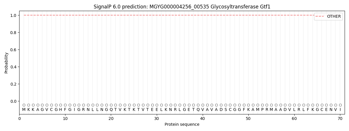
| Other | SP_Sec_SPI | LIPO_Sec_SPII | TAT_Tat_SPI | TATLIP_Sec_SPII | PILIN_Sec_SPIII |
|---|---|---|---|---|---|
| 1.000074 | 0.000000 | 0.000000 | 0.000000 | 0.000000 | 0.000000 |