You are browsing environment: HUMAN GUT
CAZyme Information: MGYG000002416_00399
You are here: Home > Sequence: MGYG000002416_00399
Basic Information |
Genomic context |
Full Sequence |
Enzyme annotations |
CAZy signature domains |
CDD domains |
CAZyme hits |
PDB hits |
Swiss-Prot hits |
SignalP and Lipop annotations |
TMHMM annotations
Basic Information help
| Species | Hydrogeniiclostridium mannosilyticum | |||||||||||
|---|---|---|---|---|---|---|---|---|---|---|---|---|
| Lineage | Bacteria; Firmicutes_A; Clostridia; Oscillospirales; Acutalibacteraceae; Hydrogeniiclostridium; Hydrogeniiclostridium mannosilyticum | |||||||||||
| CAZyme ID | MGYG000002416_00399 | |||||||||||
| CAZy Family | GH109 | |||||||||||
| CAZyme Description | Alpha-N-acetylgalactosaminidase | |||||||||||
| CAZyme Property |
|
|||||||||||
| Genome Property |
|
|||||||||||
| Gene Location | Start: 425050; End: 426198 Strand: + | |||||||||||
CAZyme Signature Domains help
| Family | Start | End | Evalue | family coverage |
|---|---|---|---|---|
| GH109 | 2 | 370 | 1.5e-97 | 0.9699248120300752 |
CDD Domains download full data without filtering help
| Cdd ID | Domain | E-Value | qStart | qEnd | sStart | sEnd | Domain Description |
|---|---|---|---|---|---|---|---|
| COG0673 | MviM | 7.53e-35 | 1 | 366 | 3 | 341 | Predicted dehydrogenase [General function prediction only]. |
| pfam01408 | GFO_IDH_MocA | 9.90e-13 | 2 | 115 | 1 | 117 | Oxidoreductase family, NAD-binding Rossmann fold. This family of enzymes utilize NADP or NAD. This family is called the GFO/IDH/MOCA family in swiss-prot. |
| PRK11579 | PRK11579 | 1.61e-05 | 53 | 113 | 60 | 117 | putative oxidoreductase; Provisional |
| smart00859 | Semialdhyde_dh | 3.45e-04 | 3 | 89 | 1 | 97 | Semialdehyde dehydrogenase, NAD binding domain. The semialdehyde dehydrogenase family is found in N-acetyl-glutamine semialdehyde dehydrogenase (AgrC), which is involved in arginine biosynthesis, and aspartate-semialdehyde dehydrogenase, an enzyme involved in the biosynthesis of various amino acids from aspartate. This family is also found in yeast and fungal Arg5,6 protein, which is cleaved into the enzymes N-acety-gamma-glutamyl-phosphate reductase and acetylglutamate kinase. These are also involved in arginine biosynthesis. All proteins in this entry contain a NAD binding region of semialdehyde dehydrogenase. |
| smart00881 | CoA_binding | 0.009 | 4 | 84 | 11 | 89 | CoA binding domain. This domain has a Rossmann fold and is found in a number of proteins including succinyl CoA synthetases, malate and ATP-citrate ligases. |
CAZyme Hits help
| Hit ID | E-Value | Query Start | Query End | Hit Start | Hit End |
|---|---|---|---|---|---|
| QHW35532.1 | 3.92e-183 | 2 | 381 | 3 | 381 |
| QHW33651.1 | 4.46e-180 | 2 | 381 | 3 | 382 |
| QHT62446.1 | 7.35e-179 | 2 | 381 | 3 | 382 |
| BBH24596.1 | 1.62e-175 | 2 | 381 | 3 | 381 |
| AEE96970.1 | 9.97e-175 | 1 | 381 | 1 | 374 |
PDB Hits download full data without filtering help
| Hit ID | E-Value | Query Start | Query End | Hit Start | Hit End | Description |
|---|---|---|---|---|---|---|
| 2IXA_A | 4.23e-37 | 8 | 368 | 29 | 426 | A-zyme,N-acetylgalactosaminidase [Elizabethkingia meningoseptica],2IXB_A Crystal structure of N-ACETYLGALACTOSAMINIDASE in complex with GalNAC [Elizabethkingia meningoseptica] |
| 6T2B_A | 7.58e-32 | 8 | 370 | 51 | 435 | Glycosidehydrolase family 109 from Akkermansia muciniphila in complex with GalNAc and NAD+. [Akkermansia muciniphila],6T2B_B Glycoside hydrolase family 109 from Akkermansia muciniphila in complex with GalNAc and NAD+. [Akkermansia muciniphila],6T2B_C Glycoside hydrolase family 109 from Akkermansia muciniphila in complex with GalNAc and NAD+. [Akkermansia muciniphila],6T2B_D Glycoside hydrolase family 109 from Akkermansia muciniphila in complex with GalNAc and NAD+. [Akkermansia muciniphila] |
| 3MOI_A | 5.83e-13 | 2 | 139 | 3 | 143 | Thecrystal structure of the putative dehydrogenase from Bordetella bronchiseptica RB50 [Bordetella bronchiseptica] |
| 6JW6_A | 4.55e-10 | 2 | 275 | 5 | 284 | Thecrystal structure of KanD2 in complex with NAD [Streptomyces kanamyceticus],6JW6_B The crystal structure of KanD2 in complex with NAD [Streptomyces kanamyceticus],6JW7_A The crystal structure of KanD2 in complex with NADH and 3'-deamino-3'-hydroxykanamycin A [Streptomyces kanamyceticus],6JW7_B The crystal structure of KanD2 in complex with NADH and 3'-deamino-3'-hydroxykanamycin A [Streptomyces kanamyceticus],6JW8_A The crystal structure of KanD2 in complex with NADH and 3'-deamino-3'-hydroxykanamycin B [Streptomyces kanamyceticus],6JW8_B The crystal structure of KanD2 in complex with NADH and 3'-deamino-3'-hydroxykanamycin B [Streptomyces kanamyceticus] |
| 3Q2I_A | 5.42e-09 | 25 | 310 | 40 | 316 | Crystalstructure of the WlbA dehydrognase from Chromobactrium violaceum in complex with NADH and UDP-GlcNAcA at 1.50 A resolution [Chromobacterium violaceum] |
Swiss-Prot Hits download full data without filtering help
| Hit ID | E-Value | Query Start | Query End | Hit Start | Hit End | Description |
|---|---|---|---|---|---|---|
| A4FN60 | 1.03e-48 | 2 | 370 | 61 | 443 | Glycosyl hydrolase family 109 protein OS=Saccharopolyspora erythraea (strain ATCC 11635 / DSM 40517 / JCM 4748 / NBRC 13426 / NCIMB 8594 / NRRL 2338) OX=405948 GN=SACE_6314 PE=3 SV=1 |
| Q01S58 | 1.49e-44 | 2 | 370 | 43 | 429 | Glycosyl hydrolase family 109 protein OS=Solibacter usitatus (strain Ellin6076) OX=234267 GN=Acid_6590 PE=3 SV=1 |
| Q0HWR6 | 3.08e-44 | 1 | 370 | 54 | 445 | Glycosyl hydrolase family 109 protein 1 OS=Shewanella sp. (strain MR-7) OX=60481 GN=Shewmr7_1440 PE=3 SV=1 |
| Q0HKG4 | 3.08e-44 | 1 | 370 | 54 | 445 | Glycosyl hydrolase family 109 protein 1 OS=Shewanella sp. (strain MR-4) OX=60480 GN=Shewmr4_1375 PE=3 SV=1 |
| A4Y8C8 | 4.29e-44 | 1 | 370 | 54 | 445 | Glycosyl hydrolase family 109 protein OS=Shewanella putrefaciens (strain CN-32 / ATCC BAA-453) OX=319224 GN=Sputcn32_2490 PE=3 SV=1 |
SignalP and Lipop Annotations help
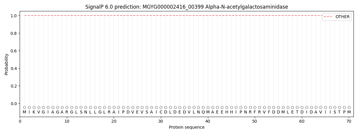
This protein is predicted as OTHER

| Other | SP_Sec_SPI | LIPO_Sec_SPII | TAT_Tat_SPI | TATLIP_Sec_SPII | PILIN_Sec_SPIII |
|---|---|---|---|---|---|
| 1.000060 | 0.000000 | 0.000000 | 0.000000 | 0.000000 | 0.000000 |
