You are browsing environment: HUMAN GUT
CAZyme Information: MGYG000002288_02189
You are here: Home > Sequence: MGYG000002288_02189
Basic Information |
Genomic context |
Full Sequence |
Enzyme annotations |
CAZy signature domains |
CDD domains |
CAZyme hits |
PDB hits |
Swiss-Prot hits |
SignalP and Lipop annotations |
TMHMM annotations
Basic Information help
| Species | Bacillus_A luti | |||||||||||
|---|---|---|---|---|---|---|---|---|---|---|---|---|
| Lineage | Bacteria; Firmicutes; Bacilli; Bacillales; Bacillaceae_G; Bacillus_A; Bacillus_A luti | |||||||||||
| CAZyme ID | MGYG000002288_02189 | |||||||||||
| CAZy Family | GT4 | |||||||||||
| CAZyme Description | GDP-mannose-dependent alpha-mannosyltransferase | |||||||||||
| CAZyme Property |
|
|||||||||||
| Genome Property |
|
|||||||||||
| Gene Location | Start: 65667; End: 66809 Strand: + | |||||||||||
CDD Domains download full data without filtering help
| Cdd ID | Domain | E-Value | qStart | qEnd | sStart | sEnd | Domain Description |
|---|---|---|---|---|---|---|---|
| cd03814 | GT4-like | 2.24e-142 | 2 | 367 | 1 | 364 | glycosyltransferase family 4 proteins. This family is most closely related to the GT4 family of glycosyltransferases and includes a sequence annotated as alpha-D-mannose-alpha(1-6)phosphatidyl myo-inositol monomannoside transferase from Bacillus halodurans. Glycosyltransferases catalyze the transfer of sugar moieties from activated donor molecules to specific acceptor molecules, forming glycosidic bonds. The acceptor molecule can be a lipid, a protein, a heterocyclic compound, or another carbohydrate residue. This group of glycosyltransferases is most closely related to the previously defined glycosyltransferase family 1 (GT1). The members of this family may transfer UDP, ADP, GDP, or CMP linked sugars. The diverse enzymatic activities among members of this family reflect a wide range of biological functions. The protein structure available for this family has the GTB topology, one of the two protein topologies observed for nucleotide-sugar-dependent glycosyltransferases. GTB proteins have distinct N- and C- terminal domains each containing a typical Rossmann fold. The two domains have high structural homology despite minimal sequence homology. The large cleft that separates the two domains includes the catalytic center and permits a high degree of flexibility. The members of this family are found mainly in bacteria and eukaryotes. |
| cd03817 | GT4_UGDG-like | 7.22e-77 | 2 | 363 | 1 | 372 | UDP-Glc:1,2-diacylglycerol 3-a-glucosyltransferase and similar proteins. This family is most closely related to the GT1 family of glycosyltransferases. UDP-glucose-diacylglycerol glucosyltransferase (EC 2.4.1.337, UGDG; also known as 1,2-diacylglycerol 3-glucosyltransferase) catalyzes the transfer of glucose from UDP-glucose to 1,2-diacylglycerol forming 3-D-glucosyl-1,2-diacylglycerol. |
| PLN02871 | PLN02871 | 5.56e-72 | 2 | 357 | 60 | 419 | UDP-sulfoquinovose:DAG sulfoquinovosyltransferase |
| COG0438 | RfaB | 4.30e-60 | 1 | 374 | 1 | 381 | Glycosyltransferase involved in cell wall bisynthesis [Cell wall/membrane/envelope biogenesis]. |
| cd03801 | GT4_PimA-like | 1.74e-55 | 2 | 359 | 1 | 357 | phosphatidyl-myo-inositol mannosyltransferase. This family is most closely related to the GT4 family of glycosyltransferases and named after PimA in Propionibacterium freudenreichii, which is involved in the biosynthesis of phosphatidyl-myo-inositol mannosides (PIM) which are early precursors in the biosynthesis of lipomannans (LM) and lipoarabinomannans (LAM), and catalyzes the addition of a mannosyl residue from GDP-D-mannose (GDP-Man) to the position 2 of the carrier lipid phosphatidyl-myo-inositol (PI) to generate a phosphatidyl-myo-inositol bearing an alpha-1,2-linked mannose residue (PIM1). Glycosyltransferases catalyze the transfer of sugar moieties from activated donor molecules to specific acceptor molecules, forming glycosidic bonds. The acceptor molecule can be a lipid, a protein, a heterocyclic compound, or another carbohydrate residue. This group of glycosyltransferases is most closely related to the previously defined glycosyltransferase family 1 (GT1). The members of this family may transfer UDP, ADP, GDP, or CMP linked sugars. The diverse enzymatic activities among members of this family reflect a wide range of biological functions. The protein structure available for this family has the GTB topology, one of the two protein topologies observed for nucleotide-sugar-dependent glycosyltransferases. GTB proteins have distinct N- and C- terminal domains each containing a typical Rossmann fold. The two domains have high structural homology despite minimal sequence homology. The large cleft that separates the two domains includes the catalytic center and permits a high degree of flexibility. The members of this family are found mainly in certain bacteria and archaea. |
CAZyme Hits help
| Hit ID | E-Value | Query Start | Query End | Hit Start | Hit End |
|---|---|---|---|---|---|
| QWH39593.1 | 7.78e-261 | 1 | 380 | 1 | 380 |
| QWH14205.1 | 6.12e-258 | 1 | 380 | 1 | 380 |
| QWG35695.1 | 6.12e-258 | 1 | 380 | 1 | 380 |
| QWG47057.1 | 6.12e-258 | 1 | 380 | 1 | 380 |
| QWG30164.1 | 2.49e-257 | 1 | 380 | 1 | 380 |
PDB Hits download full data without filtering help
| Hit ID | E-Value | Query Start | Query End | Hit Start | Hit End | Description |
|---|---|---|---|---|---|---|
| 3C4Q_A | 1.37e-16 | 174 | 377 | 196 | 412 | Structureof the retaining glycosyltransferase MshA : The first step in mycothiol biosynthesis. Organism : Corynebacterium glutamicum- Complex with UDP [Corynebacterium glutamicum],3C4Q_B Structure of the retaining glycosyltransferase MshA : The first step in mycothiol biosynthesis. Organism : Corynebacterium glutamicum- Complex with UDP [Corynebacterium glutamicum],3C4V_A Structure of the retaining glycosyltransferase MshA:The first step in mycothiol biosynthesis. Organism: Corynebacterium glutamicum : Complex with UDP and 1L-INS-1-P. [Corynebacterium glutamicum],3C4V_B Structure of the retaining glycosyltransferase MshA:The first step in mycothiol biosynthesis. Organism: Corynebacterium glutamicum : Complex with UDP and 1L-INS-1-P. [Corynebacterium glutamicum] |
| 3C48_A | 1.45e-16 | 174 | 377 | 216 | 432 | Structureof the retaining glycosyltransferase MshA: The first step in mycothiol biosynthesis. Organism: Corynebacterium glutamicum- APO (OPEN) structure. [Corynebacterium glutamicum],3C48_B Structure of the retaining glycosyltransferase MshA: The first step in mycothiol biosynthesis. Organism: Corynebacterium glutamicum- APO (OPEN) structure. [Corynebacterium glutamicum] |
| 2JJM_A | 2.25e-15 | 162 | 343 | 178 | 357 | CrystalStructure of a family GT4 glycosyltransferase from Bacillus anthracis ORF BA1558. [Bacillus anthracis str. Ames],2JJM_B Crystal Structure of a family GT4 glycosyltransferase from Bacillus anthracis ORF BA1558. [Bacillus anthracis str. Ames],2JJM_C Crystal Structure of a family GT4 glycosyltransferase from Bacillus anthracis ORF BA1558. [Bacillus anthracis str. Ames],2JJM_D Crystal Structure of a family GT4 glycosyltransferase from Bacillus anthracis ORF BA1558. [Bacillus anthracis str. Ames],2JJM_E Crystal Structure of a family GT4 glycosyltransferase from Bacillus anthracis ORF BA1558. [Bacillus anthracis str. Ames],2JJM_F Crystal Structure of a family GT4 glycosyltransferase from Bacillus anthracis ORF BA1558. [Bacillus anthracis str. Ames],2JJM_G Crystal Structure of a family GT4 glycosyltransferase from Bacillus anthracis ORF BA1558. [Bacillus anthracis str. Ames],2JJM_H Crystal Structure of a family GT4 glycosyltransferase from Bacillus anthracis ORF BA1558. [Bacillus anthracis str. Ames],2JJM_I Crystal Structure of a family GT4 glycosyltransferase from Bacillus anthracis ORF BA1558. [Bacillus anthracis str. Ames],2JJM_J Crystal Structure of a family GT4 glycosyltransferase from Bacillus anthracis ORF BA1558. [Bacillus anthracis str. Ames],2JJM_K Crystal Structure of a family GT4 glycosyltransferase from Bacillus anthracis ORF BA1558. [Bacillus anthracis str. Ames],2JJM_L Crystal Structure of a family GT4 glycosyltransferase from Bacillus anthracis ORF BA1558. [Bacillus anthracis str. Ames] |
| 3MBO_A | 2.48e-15 | 162 | 343 | 198 | 377 | CrystalStructure of the Glycosyltransferase BaBshA bound with UDP and L-malate [Bacillus anthracis],3MBO_B Crystal Structure of the Glycosyltransferase BaBshA bound with UDP and L-malate [Bacillus anthracis],3MBO_C Crystal Structure of the Glycosyltransferase BaBshA bound with UDP and L-malate [Bacillus anthracis],3MBO_D Crystal Structure of the Glycosyltransferase BaBshA bound with UDP and L-malate [Bacillus anthracis],3MBO_E Crystal Structure of the Glycosyltransferase BaBshA bound with UDP and L-malate [Bacillus anthracis],3MBO_F Crystal Structure of the Glycosyltransferase BaBshA bound with UDP and L-malate [Bacillus anthracis],3MBO_G Crystal Structure of the Glycosyltransferase BaBshA bound with UDP and L-malate [Bacillus anthracis],3MBO_H Crystal Structure of the Glycosyltransferase BaBshA bound with UDP and L-malate [Bacillus anthracis] |
| 6KIH_A | 2.98e-14 | 174 | 334 | 222 | 387 | Sucrose-phosphatesynthase (tll1590) from Thermosynechococcus elongatus [Thermosynechococcus vestitus],6KIH_B Sucrose-phosphate synthase (tll1590) from Thermosynechococcus elongatus [Thermosynechococcus vestitus],6KIH_C Sucrose-phosphate synthase (tll1590) from Thermosynechococcus elongatus [Thermosynechococcus vestitus],6KIH_D Sucrose-phosphate synthase (tll1590) from Thermosynechococcus elongatus [Thermosynechococcus vestitus],6KIH_E Sucrose-phosphate synthase (tll1590) from Thermosynechococcus elongatus [Thermosynechococcus vestitus],6KIH_F Sucrose-phosphate synthase (tll1590) from Thermosynechococcus elongatus [Thermosynechococcus vestitus],6KIH_G Sucrose-phosphate synthase (tll1590) from Thermosynechococcus elongatus [Thermosynechococcus vestitus],6KIH_H Sucrose-phosphate synthase (tll1590) from Thermosynechococcus elongatus [Thermosynechococcus vestitus],6KIH_I Sucrose-phosphate synthase (tll1590) from Thermosynechococcus elongatus [Thermosynechococcus vestitus],6KIH_J Sucrose-phosphate synthase (tll1590) from Thermosynechococcus elongatus [Thermosynechococcus vestitus],6KIH_K Sucrose-phosphate synthase (tll1590) from Thermosynechococcus elongatus [Thermosynechococcus vestitus],6KIH_L Sucrose-phosphate synthase (tll1590) from Thermosynechococcus elongatus [Thermosynechococcus vestitus] |
Swiss-Prot Hits download full data without filtering help
| Hit ID | E-Value | Query Start | Query End | Hit Start | Hit End | Description |
|---|---|---|---|---|---|---|
| P9WMY5 | 3.73e-57 | 1 | 377 | 4 | 376 | GDP-mannose-dependent alpha-mannosyltransferase OS=Mycobacterium tuberculosis (strain ATCC 25618 / H37Rv) OX=83332 GN=mgtA PE=1 SV=1 |
| P9WMY4 | 3.73e-57 | 1 | 377 | 4 | 376 | GDP-mannose-dependent alpha-mannosyltransferase OS=Mycobacterium tuberculosis (strain CDC 1551 / Oshkosh) OX=83331 GN=mgtA PE=3 SV=1 |
| Q8NT41 | 4.52e-57 | 1 | 371 | 7 | 375 | GDP-mannose-dependent alpha-mannosyltransferase OS=Corynebacterium glutamicum (strain ATCC 13032 / DSM 20300 / BCRC 11384 / JCM 1318 / LMG 3730 / NCIMB 10025) OX=196627 GN=mgtA PE=1 SV=1 |
| A0QRG8 | 7.00e-52 | 1 | 375 | 1 | 371 | GDP-mannose-dependent alpha-mannosyltransferase OS=Mycolicibacterium smegmatis (strain ATCC 700084 / mc(2)155) OX=246196 GN=mgtA PE=3 SV=1 |
| Q8S4F6 | 9.77e-46 | 2 | 356 | 105 | 465 | Sulfoquinovosyl transferase SQD2 OS=Arabidopsis thaliana OX=3702 GN=SQD2 PE=1 SV=1 |
SignalP and Lipop Annotations help
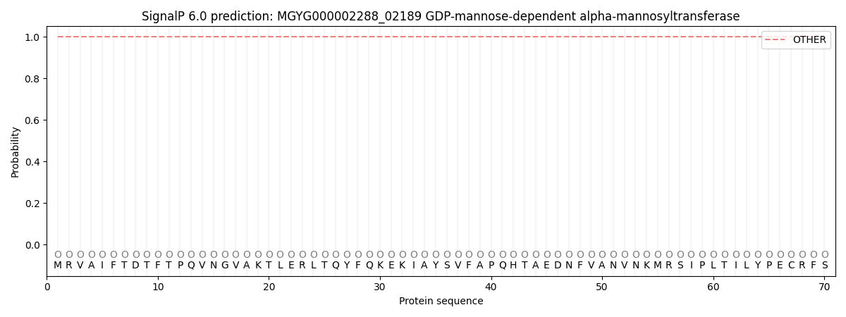
This protein is predicted as OTHER
| Other | SP_Sec_SPI | LIPO_Sec_SPII | TAT_Tat_SPI | TATLIP_Sec_SPII | PILIN_Sec_SPIII |
|---|---|---|---|---|---|
| 1.000043 | 0.000000 | 0.000000 | 0.000000 | 0.000000 | 0.000000 |