

You are browsing environment: HUMAN GUT
CAZyme Information: MGYG000002061_01889
You are here: Home > Sequence: MGYG000002061_01889
Basic Information |
Genomic context |
Full Sequence |
Enzyme annotations |
CAZy signature domains |
CDD domains |
CAZyme hits |
PDB hits |
Swiss-Prot hits |
SignalP and Lipop annotations |
TMHMM annotations
Basic Information help
| Species | QAMX01 sp004555605 | |||||||||||
|---|---|---|---|---|---|---|---|---|---|---|---|---|
| Lineage | Bacteria; Firmicutes_A; Clostridia; Oscillospirales; QAMX01; QAMX01; QAMX01 sp004555605 | |||||||||||
| CAZyme ID | MGYG000002061_01889 | |||||||||||
| CAZy Family | GH109 | |||||||||||
| CAZyme Description | Alpha-N-acetylgalactosaminidase | |||||||||||
| CAZyme Property |
|
|||||||||||
| Genome Property |
|
|||||||||||
| Gene Location | Start: 2379; End: 3608 Strand: - | |||||||||||
CAZyme Signature Domains help
| Family | Start | End | Evalue | family coverage |
|---|---|---|---|---|
| GH109 | 3 | 389 | 6.6e-150 | 0.9849624060150376 |
CDD Domains download full data without filtering help
| Cdd ID | Domain | E-Value | qStart | qEnd | sStart | sEnd | Domain Description |
|---|---|---|---|---|---|---|---|
| COG0673 | MviM | 6.69e-21 | 3 | 163 | 1 | 158 | Predicted dehydrogenase [General function prediction only]. |
| pfam01408 | GFO_IDH_MocA | 8.35e-11 | 6 | 127 | 1 | 117 | Oxidoreductase family, NAD-binding Rossmann fold. This family of enzymes utilize NADP or NAD. This family is called the GFO/IDH/MOCA family in swiss-prot. |
CAZyme Hits help
| Hit ID | E-Value | Query Start | Query End | Hit Start | Hit End |
|---|---|---|---|---|---|
| AEE95391.1 | 2.66e-184 | 2 | 409 | 5 | 412 |
| QTH41090.1 | 1.23e-173 | 6 | 390 | 10 | 394 |
| ANY74959.1 | 6.32e-167 | 5 | 393 | 11 | 399 |
| AEF80535.1 | 6.76e-157 | 2 | 405 | 5 | 413 |
| ALS29040.1 | 1.28e-155 | 1 | 396 | 1 | 402 |
PDB Hits download full data without filtering help
| Hit ID | E-Value | Query Start | Query End | Hit Start | Hit End | Description |
|---|---|---|---|---|---|---|
| 2IXA_A | 2.01e-103 | 6 | 387 | 21 | 429 | A-zyme,N-acetylgalactosaminidase [Elizabethkingia meningoseptica],2IXB_A Crystal structure of N-ACETYLGALACTOSAMINIDASE in complex with GalNAC [Elizabethkingia meningoseptica] |
| 6T2B_A | 6.23e-87 | 6 | 394 | 43 | 443 | Glycosidehydrolase family 109 from Akkermansia muciniphila in complex with GalNAc and NAD+. [Akkermansia muciniphila],6T2B_B Glycoside hydrolase family 109 from Akkermansia muciniphila in complex with GalNAc and NAD+. [Akkermansia muciniphila],6T2B_C Glycoside hydrolase family 109 from Akkermansia muciniphila in complex with GalNAc and NAD+. [Akkermansia muciniphila],6T2B_D Glycoside hydrolase family 109 from Akkermansia muciniphila in complex with GalNAc and NAD+. [Akkermansia muciniphila] |
| 3E18_A | 4.50e-13 | 1 | 155 | 1 | 148 | CRYSTALSTRUCTURE OF NAD-BINDING PROTEIN FROM Listeria innocua [Listeria innocua],3E18_B CRYSTAL STRUCTURE OF NAD-BINDING PROTEIN FROM Listeria innocua [Listeria innocua] |
| 3EC7_A | 4.72e-10 | 2 | 152 | 20 | 168 | CrystalStructure of Putative Dehydrogenase from Salmonella typhimurium LT2 [Salmonella enterica subsp. enterica serovar Typhimurium str. LT2],3EC7_B Crystal Structure of Putative Dehydrogenase from Salmonella typhimurium LT2 [Salmonella enterica subsp. enterica serovar Typhimurium str. LT2],3EC7_C Crystal Structure of Putative Dehydrogenase from Salmonella typhimurium LT2 [Salmonella enterica subsp. enterica serovar Typhimurium str. LT2],3EC7_D Crystal Structure of Putative Dehydrogenase from Salmonella typhimurium LT2 [Salmonella enterica subsp. enterica serovar Typhimurium str. LT2],3EC7_E Crystal Structure of Putative Dehydrogenase from Salmonella typhimurium LT2 [Salmonella enterica subsp. enterica serovar Typhimurium str. LT2],3EC7_F Crystal Structure of Putative Dehydrogenase from Salmonella typhimurium LT2 [Salmonella enterica subsp. enterica serovar Typhimurium str. LT2],3EC7_G Crystal Structure of Putative Dehydrogenase from Salmonella typhimurium LT2 [Salmonella enterica subsp. enterica serovar Typhimurium str. LT2],3EC7_H Crystal Structure of Putative Dehydrogenase from Salmonella typhimurium LT2 [Salmonella enterica subsp. enterica serovar Typhimurium str. LT2] |
| 4FB5_A | 3.82e-07 | 11 | 163 | 28 | 185 | Crystalstructure of a probable oxidoreduxtase protein [Rhizobium etli CFN 42],4FB5_B Crystal structure of a probable oxidoreduxtase protein [Rhizobium etli CFN 42] |
Swiss-Prot Hits download full data without filtering help
| Hit ID | E-Value | Query Start | Query End | Hit Start | Hit End | Description |
|---|---|---|---|---|---|---|
| B2FLK4 | 2.19e-113 | 2 | 394 | 30 | 446 | Glycosyl hydrolase family 109 protein OS=Stenotrophomonas maltophilia (strain K279a) OX=522373 GN=Smlt4431 PE=3 SV=1 |
| A4Q8G1 | 1.22e-110 | 11 | 394 | 58 | 464 | Alpha-N-acetylgalactosaminidase OS=Tannerella forsythia OX=28112 GN=nagA PE=3 SV=1 |
| A6LB54 | 1.32e-106 | 11 | 395 | 55 | 463 | Glycosyl hydrolase family 109 protein OS=Parabacteroides distasonis (strain ATCC 8503 / DSM 20701 / CIP 104284 / JCM 5825 / NCTC 11152) OX=435591 GN=BDI_1155 PE=3 SV=1 |
| A4Q8F7 | 1.10e-102 | 6 | 387 | 21 | 429 | Alpha-N-acetylgalactosaminidase OS=Elizabethkingia meningoseptica OX=238 GN=nagA PE=1 SV=1 |
| Q01S58 | 2.09e-98 | 3 | 394 | 40 | 437 | Glycosyl hydrolase family 109 protein OS=Solibacter usitatus (strain Ellin6076) OX=234267 GN=Acid_6590 PE=3 SV=1 |
SignalP and Lipop Annotations help
This protein is predicted as OTHER
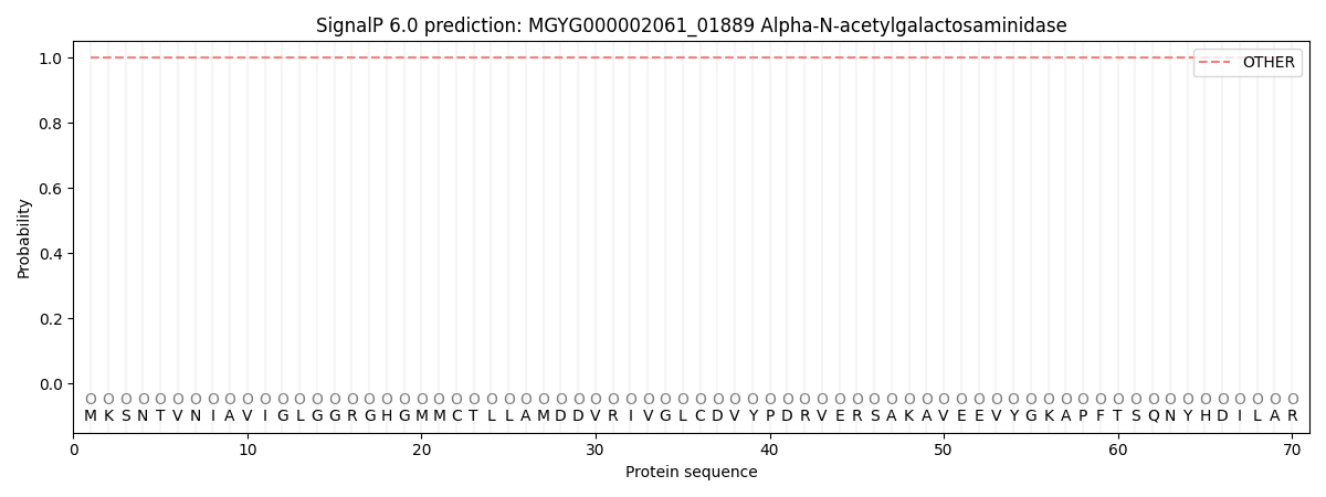
| Other | SP_Sec_SPI | LIPO_Sec_SPII | TAT_Tat_SPI | TATLIP_Sec_SPII | PILIN_Sec_SPIII |
|---|---|---|---|---|---|
| 1.000061 | 0.000001 | 0.000000 | 0.000000 | 0.000000 | 0.000000 |