You are browsing environment: HUMAN GUT
CAZyme Information: MGYG000001905_00263
You are here: Home > Sequence: MGYG000001905_00263
Basic Information |
Genomic context |
Full Sequence |
Enzyme annotations |
CAZy signature domains |
CDD domains |
CAZyme hits |
PDB hits |
Swiss-Prot hits |
SignalP and Lipop annotations |
TMHMM annotations
Basic Information help
| Species | ||||||||||||
|---|---|---|---|---|---|---|---|---|---|---|---|---|
| Lineage | Bacteria; Firmicutes_A; Clostridia; Oscillospirales; CAG-272; Firm-07; | |||||||||||
| CAZyme ID | MGYG000001905_00263 | |||||||||||
| CAZy Family | GH4 | |||||||||||
| CAZyme Description | Alpha-galactosidase | |||||||||||
| CAZyme Property |
|
|||||||||||
| Genome Property |
|
|||||||||||
| Gene Location | Start: 117628; End: 118953 Strand: + | |||||||||||
CAZyme Signature Domains help
| Family | Start | End | Evalue | family coverage |
|---|---|---|---|---|
| GH4 | 3 | 185 | 3.8e-66 | 0.9832402234636871 |
CDD Domains download full data without filtering help
| Cdd ID | Domain | E-Value | qStart | qEnd | sStart | sEnd | Domain Description |
|---|---|---|---|---|---|---|---|
| PRK15076 | PRK15076 | 0.0 | 1 | 441 | 1 | 431 | alpha-galactosidase; Provisional |
| cd05297 | GH4_alpha_glucosidase_galactosidase | 0.0 | 3 | 430 | 2 | 423 | Glycoside Hydrolases Family 4; Alpha-glucosidases and alpha-galactosidases. linked to 3D####ucture |
| COG1486 | CelF | 2.83e-169 | 1 | 441 | 3 | 440 | Alpha-galactosidase/6-phospho-beta-glucosidase, family 4 of glycosyl hydrolase [Carbohydrate transport and metabolism]. |
| pfam02056 | Glyco_hydro_4 | 1.95e-73 | 3 | 187 | 1 | 181 | Family 4 glycosyl hydrolase. |
| cd05296 | GH4_P_beta_glucosidase | 4.25e-68 | 3 | 437 | 2 | 419 | Glycoside Hydrolases Family 4; Phospho-beta-glucosidase. Some bacteria simultaneously translocate and phosphorylate disaccharides via the phosphoenolpyruvate-dependent phosphotransferase system (PEP-PTS). After translocation, these phospho-disaccharides may be hydrolyzed by the GH4 glycoside hydrolases such as the phospho-beta-glucosidases. Other organisms (such as archaea and Thermotoga maritima ) lack the PEP-PTS system, but have several enzymes normally associated with the PEP-PTS operon. The 6-phospho-beta-glucosidase from Thermotoga maritima hydrolylzes cellobiose 6-phosphate (6P) into glucose-6P and glucose, in an NAD+ and Mn2+ dependent fashion. The Escherichia coli 6-phospho-beta-glucosidase (also called celF) hydrolyzes a variety of phospho-beta-glucosides including cellobiose-6P, salicin-6P, arbutin-6P, and gentobiose-6P. Phospho-beta-glucosidases are part of the NAD(P)-binding Rossmann fold superfamily, which includes a wide variety of protein families including the NAD(P)-binding domains of alcohol dehydrogenases, tyrosine-dependent oxidoreductases, glyceraldehyde-3-phosphate dehydrogenases, formate/glycerate dehydrogenases, siroheme synthases, 6-phosphogluconate dehydrogenases, aminoacid dehydrogenases, repressor rex, and NAD-binding potassium channel domains, among others. |
CAZyme Hits help
| Hit ID | E-Value | Query Start | Query End | Hit Start | Hit End |
|---|---|---|---|---|---|
| ACL77714.1 | 1.58e-219 | 1 | 440 | 1 | 440 |
| QBE95109.1 | 3.06e-219 | 1 | 440 | 1 | 439 |
| QIB56181.1 | 6.17e-219 | 1 | 440 | 1 | 439 |
| QMW81085.1 | 6.17e-219 | 1 | 440 | 1 | 439 |
| AEY68140.1 | 6.07e-217 | 1 | 440 | 1 | 440 |
PDB Hits download full data without filtering help
| Hit ID | E-Value | Query Start | Query End | Hit Start | Hit End | Description |
|---|---|---|---|---|---|---|
| 5C3M_A | 9.91e-36 | 3 | 440 | 6 | 437 | Crystalstructure of Gan4C, a GH4 6-phospho-glucosidase from Geobacillus stearothermophilus [Geobacillus stearothermophilus],5C3M_B Crystal structure of Gan4C, a GH4 6-phospho-glucosidase from Geobacillus stearothermophilus [Geobacillus stearothermophilus],5C3M_C Crystal structure of Gan4C, a GH4 6-phospho-glucosidase from Geobacillus stearothermophilus [Geobacillus stearothermophilus],5C3M_D Crystal structure of Gan4C, a GH4 6-phospho-glucosidase from Geobacillus stearothermophilus [Geobacillus stearothermophilus] |
| 1S6Y_A | 4.07e-32 | 3 | 440 | 9 | 440 | 2.3Acrystal structure of phospho-beta-glucosidase [Geobacillus stearothermophilus] |
| 3FEF_A | 2.70e-31 | 3 | 421 | 7 | 429 | Crystalstructure of putative glucosidase lplD from bacillus subtilis [Bacillus subtilis],3FEF_B Crystal structure of putative glucosidase lplD from bacillus subtilis [Bacillus subtilis],3FEF_C Crystal structure of putative glucosidase lplD from bacillus subtilis [Bacillus subtilis],3FEF_D Crystal structure of putative glucosidase lplD from bacillus subtilis [Bacillus subtilis] |
| 1U8X_X | 3.30e-28 | 4 | 438 | 31 | 462 | CrystalStructure Of Glva From Bacillus Subtilis, A Metal-requiring, Nad-dependent 6-phospho-alpha-glucosidase [Bacillus subtilis] |
| 6DUX_A | 4.65e-26 | 76 | 441 | 77 | 443 | ChainA, 6-phospho-alpha-glucosidase [Klebsiella pneumoniae],6DUX_B Chain B, 6-phospho-alpha-glucosidase [Klebsiella pneumoniae],6DVV_A Chain A, 6-phospho-alpha-glucosidase [Klebsiella pneumoniae],6DVV_B Chain B, 6-phospho-alpha-glucosidase [Klebsiella pneumoniae] |
Swiss-Prot Hits download full data without filtering help
| Hit ID | E-Value | Query Start | Query End | Hit Start | Hit End | Description |
|---|---|---|---|---|---|---|
| O34645 | 2.00e-190 | 1 | 440 | 1 | 431 | Alpha-galactosidase OS=Bacillus subtilis (strain 168) OX=224308 GN=melA PE=1 SV=1 |
| P06720 | 9.95e-136 | 3 | 440 | 6 | 448 | Alpha-galactosidase OS=Escherichia coli (strain K12) OX=83333 GN=melA PE=1 SV=1 |
| P30877 | 2.00e-135 | 3 | 440 | 6 | 448 | Alpha-galactosidase OS=Salmonella typhimurium (strain LT2 / SGSC1412 / ATCC 700720) OX=99287 GN=melA PE=3 SV=2 |
| Q9X4Y0 | 9.82e-68 | 3 | 440 | 5 | 442 | Alpha-galactosidase OS=Rhizobium meliloti (strain 1021) OX=266834 GN=melA PE=3 SV=1 |
| Q9AI65 | 1.08e-45 | 3 | 437 | 4 | 449 | Alpha-glucosidase OS=Erwinia rhapontici OX=55212 GN=palH PE=1 SV=2 |
SignalP and Lipop Annotations help
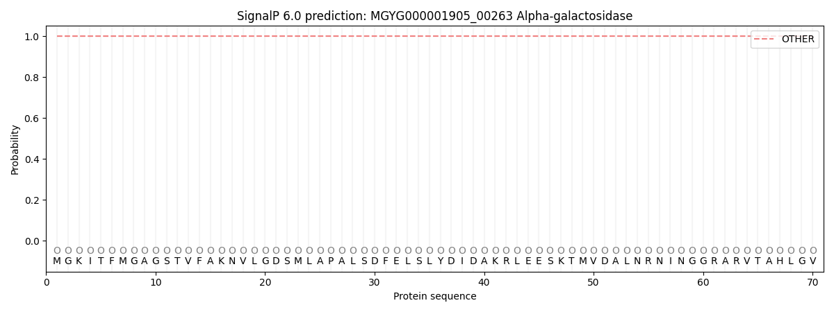
This protein is predicted as OTHER

| Other | SP_Sec_SPI | LIPO_Sec_SPII | TAT_Tat_SPI | TATLIP_Sec_SPII | PILIN_Sec_SPIII |
|---|---|---|---|---|---|
| 1.000054 | 0.000000 | 0.000000 | 0.000000 | 0.000000 | 0.000000 |
