You are browsing environment: HUMAN GUT
CAZyme Information: MGYG000002554_01153
You are here: Home > Sequence: MGYG000002554_01153
Basic Information |
Genomic context |
Full Sequence |
Enzyme annotations |
CAZy signature domains |
CDD domains |
CAZyme hits |
PDB hits |
Swiss-Prot hits |
SignalP and Lipop annotations |
TMHMM annotations
Basic Information help
| Species | Streptococcus oralis_W | |||||||||||
|---|---|---|---|---|---|---|---|---|---|---|---|---|
| Lineage | Bacteria; Firmicutes; Bacilli; Lactobacillales; Streptococcaceae; Streptococcus; Streptococcus oralis_W | |||||||||||
| CAZyme ID | MGYG000002554_01153 | |||||||||||
| CAZy Family | CBM40 | |||||||||||
| CAZyme Description | Sialidase A | |||||||||||
| CAZyme Property |
|
|||||||||||
| Genome Property |
|
|||||||||||
| Gene Location | Start: 19; End: 1050 Strand: + | |||||||||||
CDD Domains download full data without filtering help
| Cdd ID | Domain | E-Value | qStart | qEnd | sStart | sEnd | Domain Description |
|---|---|---|---|---|---|---|---|
| pfam00746 | Gram_pos_anchor | 9.57e-07 | 303 | 343 | 1 | 41 | LPXTG cell wall anchor motif. |
| TIGR01167 | LPXTG_anchor | 5.30e-06 | 310 | 343 | 1 | 34 | LPXTG-motif cell wall anchor domain. This model describes the LPXTG motif-containing region found at the C-terminus of many surface proteins of Streptococcus and Streptomyces species. Cleavage between the Thr and Gly by sortase or a related enzyme leads to covalent anchoring at the new C-terminal Thr to the cell wall. Hits that do not lie at the C-terminus or are not found in Gram-positive bacteria are probably false-positive. A common feature of this proteins containing this domain appears to be a high proportion of charged and zwitterionic residues immediatedly upstream of the LPXTG motif. This model differs from other descriptions of the LPXTG region by including a portion of that upstream charged region. [Cell envelope, Other] |
| cd15482 | Sialidase_non-viral | 5.29e-05 | 2 | 58 | 283 | 339 | Non-viral sialidases. Sialidases or neuraminidases function to bind and hydrolyze terminal sialic acid residues from various glycoconjugates, they play vital roles in pathogenesis, bacterial nutrition and cellular interactions. They have a six-bladed, beta-propeller fold with the non-viral sialidases containing 2-5 Asp-box motifs (most commonly Ser/Thr-X-Asp-[X]-Gly-X-Thr- Trp/Phe). This CD includes eubacterial and eukaryotic sialidases. |
CAZyme Hits help
| Hit ID | E-Value | Query Start | Query End | Hit Start | Hit End |
|---|---|---|---|---|---|
| QXW61877.1 | 1.12e-155 | 1 | 343 | 751 | 1093 |
| ANR74557.1 | 3.21e-149 | 1 | 343 | 753 | 1132 |
| ATF57787.1 | 9.95e-143 | 1 | 343 | 751 | 1130 |
| QLL97088.1 | 1.22e-139 | 1 | 343 | 751 | 1130 |
| APU52327.1 | 1.22e-139 | 1 | 343 | 751 | 1130 |
PDB Hits download full data without filtering help
| Hit ID | E-Value | Query Start | Query End | Hit Start | Hit End | Description |
|---|---|---|---|---|---|---|
| 2VVZ_A | 4.48e-33 | 1 | 95 | 410 | 504 | Structureof the catalytic domain of Streptococcus pneumoniae sialidase NanA [Streptococcus pneumoniae],2VVZ_B Structure of the catalytic domain of Streptococcus pneumoniae sialidase NanA [Streptococcus pneumoniae] |
| 3H71_A | 4.85e-22 | 1 | 65 | 412 | 476 | Crystalstructure of Streptococcus pneumoniae D39 neuraminidase A precursor (NanA) [Streptococcus pneumoniae R6],3H71_B Crystal structure of Streptococcus pneumoniae D39 neuraminidase A precursor (NanA) [Streptococcus pneumoniae R6] |
| 3H72_A | 4.85e-22 | 1 | 65 | 412 | 476 | Crystalstructure of Streptococcus pneumoniae D39 neuraminidase A precursor (NanA) in complex with NANA [Streptococcus pneumoniae R6],3H72_B Crystal structure of Streptococcus pneumoniae D39 neuraminidase A precursor (NanA) in complex with NANA [Streptococcus pneumoniae R6],3H73_A Crystal structure of Streptococcus pneumoniae D39 neuraminidase A precursor (NanA) in complex with DANA [Streptococcus pneumoniae R6],3H73_B Crystal structure of Streptococcus pneumoniae D39 neuraminidase A precursor (NanA) in complex with DANA [Streptococcus pneumoniae R6] |
| 6QZH_A | 8.38e-22 | 1 | 78 | 617 | 694 | Structureof the human CC Chemokine Receptor 7 in complex with the intracellular allosteric antagonist Cmp2105 and the insertion protein Sialidase NanA [Homo sapiens] |
| 2W20_A | 8.66e-22 | 1 | 64 | 408 | 471 | Structureof the catalytic domain of the native NanA sialidase from Streptococcus pneumoniae [Streptococcus pneumoniae R6],2W20_B Structure of the catalytic domain of the native NanA sialidase from Streptococcus pneumoniae [Streptococcus pneumoniae R6] |
Swiss-Prot Hits download full data without filtering help
| Hit ID | E-Value | Query Start | Query End | Hit Start | Hit End | Description |
|---|---|---|---|---|---|---|
| P62575 | 4.36e-90 | 1 | 343 | 728 | 1035 | Sialidase A OS=Streptococcus pneumoniae OX=1313 GN=nanA PE=1 SV=1 |
| P62576 | 4.36e-90 | 1 | 343 | 728 | 1035 | Sialidase A OS=Streptococcus pneumoniae (strain ATCC BAA-255 / R6) OX=171101 GN=nanA PE=1 SV=1 |
SignalP and Lipop Annotations help
This protein is predicted as OTHER
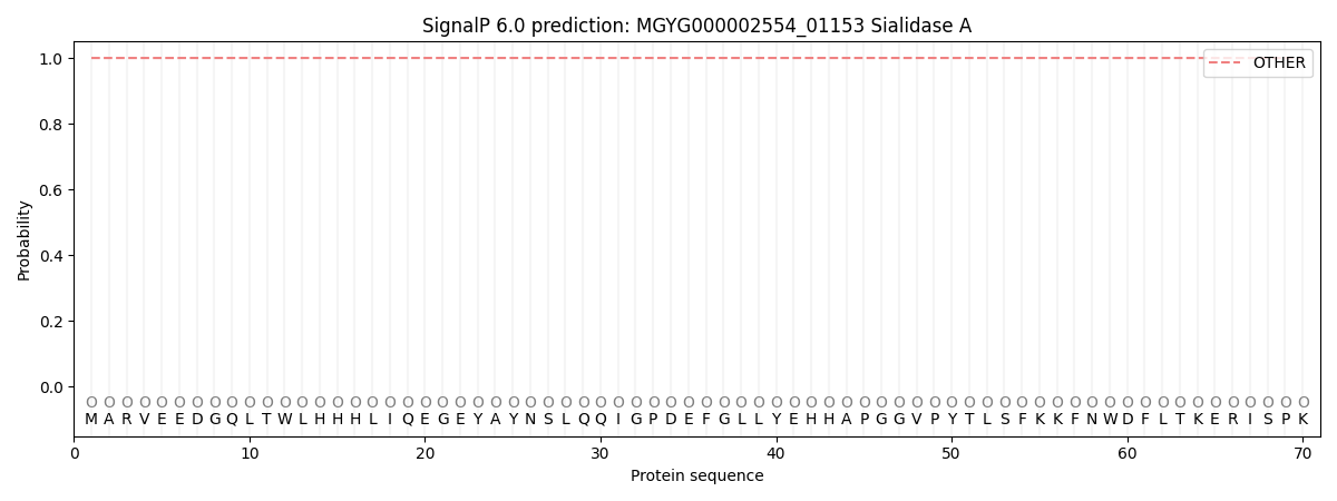
| Other | SP_Sec_SPI | LIPO_Sec_SPII | TAT_Tat_SPI | TATLIP_Sec_SPII | PILIN_Sec_SPIII |
|---|---|---|---|---|---|
| 1.000054 | 0.000001 | 0.000000 | 0.000000 | 0.000000 | 0.000000 |