You are browsing environment: HUMAN GUT
CAZyme Information: MGYG000002467_02536
You are here: Home > Sequence: MGYG000002467_02536
Basic Information |
Genomic context |
Full Sequence |
Enzyme annotations |
CAZy signature domains |
CDD domains |
CAZyme hits |
PDB hits |
Swiss-Prot hits |
SignalP and Lipop annotations |
TMHMM annotations
Basic Information help
| Species | Yersinia frederiksenii_C | |||||||||||
|---|---|---|---|---|---|---|---|---|---|---|---|---|
| Lineage | Bacteria; Proteobacteria; Gammaproteobacteria; Enterobacterales; Enterobacteriaceae; Yersinia; Yersinia frederiksenii_C | |||||||||||
| CAZyme ID | MGYG000002467_02536 | |||||||||||
| CAZy Family | GT4 | |||||||||||
| CAZyme Description | Glutamate/aspartate import solute-binding protein | |||||||||||
| CAZyme Property |
|
|||||||||||
| Genome Property |
|
|||||||||||
| Gene Location | Start: 375244; End: 376182 Strand: + | |||||||||||
CDD Domains download full data without filtering help
| Cdd ID | Domain | E-Value | qStart | qEnd | sStart | sEnd | Domain Description |
|---|---|---|---|---|---|---|---|
| PRK10797 | PRK10797 | 0.0 | 1 | 312 | 1 | 302 | glutamate and aspartate transporter subunit; Provisional |
| cd13688 | PBP2_GltI_DEBP | 2.32e-113 | 43 | 280 | 1 | 238 | Substrate-binding domain of ABC aspartate-glutamate transporter; the type 2 periplasmic binding protein fold. This subfamily represents the periplasmic-binding protein component of ABC transporter specific for carboxylic amino acids, including GtlI from Escherichia coli. The aspartate-glutamate binding domain belongs to the type 2 periplasmic binding protein fold superfamily (PBP2), whose many members are involved in chemotaxis and uptake of nutrients and other small molecules from the extracellular space as a primary receptor. The PBP2 proteins are typically comprised of two globular subdomains connected by a flexible hinge and bind their ligand in the cleft between these domains in a manner resembling a Venus flytrap. After binding their specific ligand with high affinity, they can interact with a cognate membrane transport complex comprised of two integral membrane domains and two receptor cytoplasmically-located ATPase domains. This interaction triggers the ligand translocation across the cytoplasmic membrane energized by ATP hydrolysis. |
| cd01000 | PBP2_Cys_DEBP_like | 3.03e-82 | 43 | 280 | 1 | 228 | Substrate-binding domain of cysteine- and aspartate/glutamate-binding proteins; the type 2 periplasmic-binding protein fold. This family comprises of the periplasmic-binding protein component of ABC transporters specific for cysteine and carboxylic amino acids, as well as their closely related proteins. The cysteine and aspartate-glutamate binding domains belong to the type 2 periplasmic binding protein fold superfamily (PBP2), whose many members are involved in chemotaxis and uptake of nutrients and other small molecules from the extracellular space as a primary receptor. The PBP2 proteins are typically comprised of two globular subdomains connected by a flexible hinge and bind their ligand in the cleft between these domains in a manner resembling a Venus flytrap. After binding their specific ligand with high affinity, they can interact with a cognate membrane transport complex comprised of two integral membrane domains and two receptor cytoplasmically-located ATPase domains. This interaction triggers the ligand translocation across the cytoplasmic membrane energized by ATP hydrolysis. |
| cd13689 | PBP2_BsGlnH | 1.60e-64 | 43 | 281 | 1 | 229 | Substrate binding domain of ABC glutamine transporter from Bacillus subtilis; the type 2 periplasmic-bindig protein fold. This group includes periplasmic glutamine-binding domain GlnP from Bacillus subtilis and its related proteins. The GlnP domain belongs to the type 2 periplasmic binding protein fold superfamily (PBP2), whose many members are involved in chemotaxis and uptake of nutrients and other small molecules from the extracellular space as a primary receptor. The PBP2 proteins are typically comprised of two globular subdomains connected by a flexible hinge and bind their ligand in the cleft between these domains in a manner resembling a Venus flytrap. After binding their specific ligand with high affinity, they can interact with a cognate membrane transport complex comprised of two integral membrane domains and two receptor cytoplasmically-located ATPase domains. This interaction triggers the ligand translocation across the cytoplasmic membrane energized by ATP hydrolysis. |
| pfam00497 | SBP_bac_3 | 1.28e-54 | 52 | 280 | 1 | 223 | Bacterial extracellular solute-binding proteins, family 3. |
CAZyme Hits help
| Hit ID | E-Value | Query Start | Query End | Hit Start | Hit End |
|---|---|---|---|---|---|
| QXX81055.1 | 6.16e-172 | 1 | 312 | 1 | 297 |
| QWJ92671.1 | 1.24e-171 | 1 | 312 | 1 | 297 |
| QLQ94799.1 | 1.24e-171 | 1 | 312 | 1 | 297 |
| QQE94206.1 | 1.24e-171 | 1 | 312 | 1 | 297 |
| BBV05439.1 | 3.56e-171 | 1 | 312 | 1 | 297 |
PDB Hits download full data without filtering help
| Hit ID | E-Value | Query Start | Query End | Hit Start | Hit End | Description |
|---|---|---|---|---|---|---|
| 2VHA_A | 1.28e-172 | 36 | 312 | 3 | 279 | DEBP[Shigella flexneri],2VHA_B DEBP [Shigella flexneri] |
| 2IA4_A | 3.19e-166 | 36 | 312 | 3 | 279 | Crystalstructure of Novel amino acid binding protein from Shigella flexneri [Shigella flexneri 2a str. 301],2IA4_B Crystal structure of Novel amino acid binding protein from Shigella flexneri [Shigella flexneri 2a str. 301] |
| 5EYF_A | 6.01e-28 | 37 | 282 | 2 | 238 | CrystalStructure of Solute-binding Protein from Enterococcus faecium with Bound Glutamate [Enterococcus faecium DO],5EYF_B Crystal Structure of Solute-binding Protein from Enterococcus faecium with Bound Glutamate [Enterococcus faecium DO] |
| 3K4U_A | 7.03e-14 | 50 | 280 | 5 | 227 | CRYSTALSTRUCTURE OF putative binding component of ABC transporter from Wolinella succinogenes DSM 1740 complexed with lysine [Wolinella succinogenes],3K4U_B CRYSTAL STRUCTURE OF putative binding component of ABC transporter from Wolinella succinogenes DSM 1740 complexed with lysine [Wolinella succinogenes],3K4U_C CRYSTAL STRUCTURE OF putative binding component of ABC transporter from Wolinella succinogenes DSM 1740 complexed with lysine [Wolinella succinogenes],3K4U_D CRYSTAL STRUCTURE OF putative binding component of ABC transporter from Wolinella succinogenes DSM 1740 complexed with lysine [Wolinella succinogenes],3K4U_E CRYSTAL STRUCTURE OF putative binding component of ABC transporter from Wolinella succinogenes DSM 1740 complexed with lysine [Wolinella succinogenes],3K4U_F CRYSTAL STRUCTURE OF putative binding component of ABC transporter from Wolinella succinogenes DSM 1740 complexed with lysine [Wolinella succinogenes] |
| 1XT8_A | 1.21e-13 | 26 | 281 | 16 | 265 | ChainA, putative amino-acid transporter periplasmic solute-binding protein [Campylobacter jejuni],1XT8_B Chain B, putative amino-acid transporter periplasmic solute-binding protein [Campylobacter jejuni] |
Swiss-Prot Hits download full data without filtering help
| Hit ID | E-Value | Query Start | Query End | Hit Start | Hit End | Description |
|---|---|---|---|---|---|---|
| Q9ZF60 | 9.82e-178 | 1 | 312 | 1 | 302 | Glutamate/aspartate import solute-binding protein OS=Salmonella typhimurium (strain LT2 / SGSC1412 / ATCC 700720) OX=99287 GN=gltI PE=3 SV=3 |
| P37902 | 2.20e-174 | 1 | 312 | 1 | 302 | Glutamate/aspartate import solute-binding protein OS=Escherichia coli (strain K12) OX=83333 GN=gltI PE=1 SV=2 |
| Q9I402 | 2.37e-112 | 36 | 310 | 23 | 298 | L-glutamate/L-aspartate-binding protein OS=Pseudomonas aeruginosa (strain ATCC 15692 / DSM 22644 / CIP 104116 / JCM 14847 / LMG 12228 / 1C / PRS 101 / PAO1) OX=208964 GN=PA1342 PE=1 SV=1 |
| O34563 | 6.09e-24 | 34 | 281 | 30 | 268 | ABC transporter glutamine-binding protein GlnH OS=Bacillus subtilis (strain 168) OX=224308 GN=glnH PE=2 SV=1 |
| P27676 | 1.77e-21 | 38 | 245 | 47 | 245 | Glutamine-binding protein OS=Geobacillus stearothermophilus OX=1422 GN=glnH PE=3 SV=1 |
SignalP and Lipop Annotations help
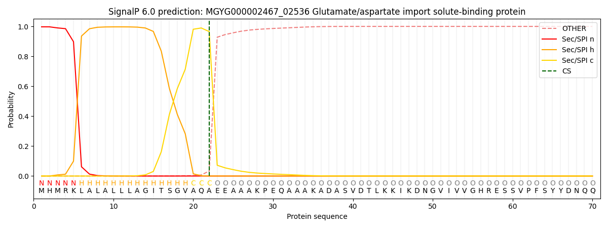
This protein is predicted as SP

| Other | SP_Sec_SPI | LIPO_Sec_SPII | TAT_Tat_SPI | TATLIP_Sec_SPII | PILIN_Sec_SPIII |
|---|---|---|---|---|---|
| 0.000625 | 0.994886 | 0.003766 | 0.000296 | 0.000216 | 0.000186 |
