You are browsing environment: HUMAN GUT
CAZyme Information: MGYG000001514_02469
You are here: Home > Sequence: MGYG000001514_02469
Basic Information |
Genomic context |
Full Sequence |
Enzyme annotations |
CAZy signature domains |
CDD domains |
CAZyme hits |
PDB hits |
Swiss-Prot hits |
SignalP and Lipop annotations |
TMHMM annotations
Basic Information help
| Species | Paenibacillus_A ihumii | |||||||||||
|---|---|---|---|---|---|---|---|---|---|---|---|---|
| Lineage | Bacteria; Firmicutes; Bacilli; Paenibacillales; Paenibacillaceae; Paenibacillus_A; Paenibacillus_A ihumii | |||||||||||
| CAZyme ID | MGYG000001514_02469 | |||||||||||
| CAZy Family | GH91 | |||||||||||
| CAZyme Description | Inulin fructotransferase [DFA-I-forming] | |||||||||||
| CAZyme Property |
|
|||||||||||
| Genome Property |
|
|||||||||||
| Gene Location | Start: 991728; End: 993071 Strand: + | |||||||||||
CAZyme Signature Domains help
| Family | Start | End | Evalue | family coverage |
|---|---|---|---|---|
| GH91 | 4 | 443 | 1.3e-197 | 0.9949367088607595 |
CDD Domains download full data without filtering help
| Cdd ID | Domain | E-Value | qStart | qEnd | sStart | sEnd | Domain Description |
|---|---|---|---|---|---|---|---|
| cd21111 | IFTase | 0.0 | 5 | 443 | 3 | 395 | inulin fructotransferase. Inulin fructotransferase (IFTase; EC 4.2.2.17 and EC 4.2.2.18), a member of the glycoside hydrolase family 91, catalyzes depolymerization of beta-2,1-fructans inulin by successively removing the terminal difructosaccharide units as cyclic anhydrides via intramolecular fructosyl transfer. As a result, IFTase produces DFA-I (alpha-D-fructofuranose-beta-D-fructofuranose 2',1:2,1'-dianhydride) and DFA-III (alpha-D-fructofuranose-beta-D-fructofuranose 2',1:2,3'-dianhydride). |
| pfam18835 | Beta_helix_2 | 3.21e-08 | 350 | 443 | 1 | 68 | Beta helix repeat of Inulin fructotransferase. This region contains a right-handed parallel beta helix repeat unit found in Inulin fructotransferase. This Pfam entry includes sequences not found by pfam13229. |
| pfam05048 | NosD | 3.32e-04 | 166 | 304 | 39 | 173 | Periplasmic copper-binding protein (NosD). NosD is a periplasmic protein which is thought to insert copper into the exported reductase apoenzyme (NosZ). This region forms a parallel beta helix domain. |
| pfam13229 | Beta_helix | 0.002 | 123 | 277 | 1 | 156 | Right handed beta helix region. This region contains a parallel beta helix region that shares some similarity with Pectate lyases. |
| pfam13229 | Beta_helix | 0.004 | 121 | 256 | 22 | 157 | Right handed beta helix region. This region contains a parallel beta helix region that shares some similarity with Pectate lyases. |
CAZyme Hits help
| Hit ID | E-Value | Query Start | Query End | Hit Start | Hit End |
|---|---|---|---|---|---|
| QIZ08397.1 | 0.0 | 1 | 447 | 1 | 447 |
| QGQ44358.1 | 0.0 | 1 | 447 | 1 | 447 |
| QGH34292.1 | 0.0 | 1 | 447 | 1 | 447 |
| AFH61120.1 | 0.0 | 1 | 447 | 1 | 447 |
| AYA74608.1 | 0.0 | 1 | 447 | 1 | 447 |
PDB Hits download full data without filtering help
| Hit ID | E-Value | Query Start | Query End | Hit Start | Hit End | Description |
|---|---|---|---|---|---|---|
| 5ZKS_A | 9.90e-248 | 1 | 446 | 1 | 444 | Crystalstructure of DFA-IIIase from Arthrobacter chlorophenolicus A6 [Pseudarthrobacter chlorophenolicus A6],5ZKU_A Crystal structure of DFA-IIIase from Arthrobacter chlorophenolicus A6 in complex with DFA-III [Pseudarthrobacter chlorophenolicus A6],5ZKU_B Crystal structure of DFA-IIIase from Arthrobacter chlorophenolicus A6 in complex with DFA-III [Pseudarthrobacter chlorophenolicus A6],5ZKU_C Crystal structure of DFA-IIIase from Arthrobacter chlorophenolicus A6 in complex with DFA-III [Pseudarthrobacter chlorophenolicus A6],5ZKU_D Crystal structure of DFA-IIIase from Arthrobacter chlorophenolicus A6 in complex with DFA-III [Pseudarthrobacter chlorophenolicus A6],5ZKU_E Crystal structure of DFA-IIIase from Arthrobacter chlorophenolicus A6 in complex with DFA-III [Pseudarthrobacter chlorophenolicus A6],5ZKU_F Crystal structure of DFA-IIIase from Arthrobacter chlorophenolicus A6 in complex with DFA-III [Pseudarthrobacter chlorophenolicus A6],5ZKW_A Crystal structure of DFA-IIIase from Arthrobacter chlorophenolicus A6 in complex with GF2 [Pseudarthrobacter chlorophenolicus A6],5ZKW_B Crystal structure of DFA-IIIase from Arthrobacter chlorophenolicus A6 in complex with GF2 [Pseudarthrobacter chlorophenolicus A6],5ZKW_C Crystal structure of DFA-IIIase from Arthrobacter chlorophenolicus A6 in complex with GF2 [Pseudarthrobacter chlorophenolicus A6],5ZKW_D Crystal structure of DFA-IIIase from Arthrobacter chlorophenolicus A6 in complex with GF2 [Pseudarthrobacter chlorophenolicus A6],5ZKW_E Crystal structure of DFA-IIIase from Arthrobacter chlorophenolicus A6 in complex with GF2 [Pseudarthrobacter chlorophenolicus A6],5ZKW_F Crystal structure of DFA-IIIase from Arthrobacter chlorophenolicus A6 in complex with GF2 [Pseudarthrobacter chlorophenolicus A6] |
| 5ZL5_A | 2.32e-246 | 1 | 446 | 1 | 444 | Crystalstructure of DFA-IIIase mutant C387A from Arthrobacter chlorophenolicus A6 [Pseudarthrobacter chlorophenolicus A6],5ZL5_B Crystal structure of DFA-IIIase mutant C387A from Arthrobacter chlorophenolicus A6 [Pseudarthrobacter chlorophenolicus A6],5ZL5_C Crystal structure of DFA-IIIase mutant C387A from Arthrobacter chlorophenolicus A6 [Pseudarthrobacter chlorophenolicus A6],5ZL5_D Crystal structure of DFA-IIIase mutant C387A from Arthrobacter chlorophenolicus A6 [Pseudarthrobacter chlorophenolicus A6],5ZL5_E Crystal structure of DFA-IIIase mutant C387A from Arthrobacter chlorophenolicus A6 [Pseudarthrobacter chlorophenolicus A6],5ZL5_F Crystal structure of DFA-IIIase mutant C387A from Arthrobacter chlorophenolicus A6 [Pseudarthrobacter chlorophenolicus A6],5ZLA_A Crystal structure of mutant C387A of DFA-IIIase from Arthrobacter chlorophenolicus A6 in complex with DFA-III [Pseudarthrobacter chlorophenolicus A6],5ZLA_B Crystal structure of mutant C387A of DFA-IIIase from Arthrobacter chlorophenolicus A6 in complex with DFA-III [Pseudarthrobacter chlorophenolicus A6],5ZLA_C Crystal structure of mutant C387A of DFA-IIIase from Arthrobacter chlorophenolicus A6 in complex with DFA-III [Pseudarthrobacter chlorophenolicus A6],5ZLA_D Crystal structure of mutant C387A of DFA-IIIase from Arthrobacter chlorophenolicus A6 in complex with DFA-III [Pseudarthrobacter chlorophenolicus A6],5ZLA_E Crystal structure of mutant C387A of DFA-IIIase from Arthrobacter chlorophenolicus A6 in complex with DFA-III [Pseudarthrobacter chlorophenolicus A6],5ZLA_F Crystal structure of mutant C387A of DFA-IIIase from Arthrobacter chlorophenolicus A6 in complex with DFA-III [Pseudarthrobacter chlorophenolicus A6] |
| 5ZKY_A | 2.06e-229 | 1 | 446 | 1 | 419 | Crystalstructure of DFA-IIIase from Arthrobacter chlorophenolicus A6 without its lid [Pseudarthrobacter chlorophenolicus A6],5ZL4_A Crystal structure of DFA-IIIase from Arthrobacter chlorophenolicus A6 wihout its lid in complex with GF2 [Pseudarthrobacter chlorophenolicus A6] |
| 2INU_A | 1.99e-116 | 1 | 377 | 9 | 370 | Crystalstructure of Inulin fructotransferase in the absence of substrate [Bacillus sp. snu-7],2INU_B Crystal structure of Inulin fructotransferase in the absence of substrate [Bacillus sp. snu-7],2INU_C Crystal structure of Inulin fructotransferase in the absence of substrate [Bacillus sp. snu-7],2INV_A Crystal structure of Inulin fructotransferase in the presence of di-fructose [Bacillus sp. snu-7],2INV_B Crystal structure of Inulin fructotransferase in the presence of di-fructose [Bacillus sp. snu-7],2INV_C Crystal structure of Inulin fructotransferase in the presence of di-fructose [Bacillus sp. snu-7] |
Swiss-Prot Hits download full data without filtering help
| Hit ID | E-Value | Query Start | Query End | Hit Start | Hit End | Description |
|---|---|---|---|---|---|---|
| P19870 | 1.82e-98 | 7 | 391 | 6 | 368 | Inulin fructotransferase [DFA-I-forming] OS=Arthrobacter globiformis OX=1665 PE=1 SV=3 |
SignalP and Lipop Annotations help
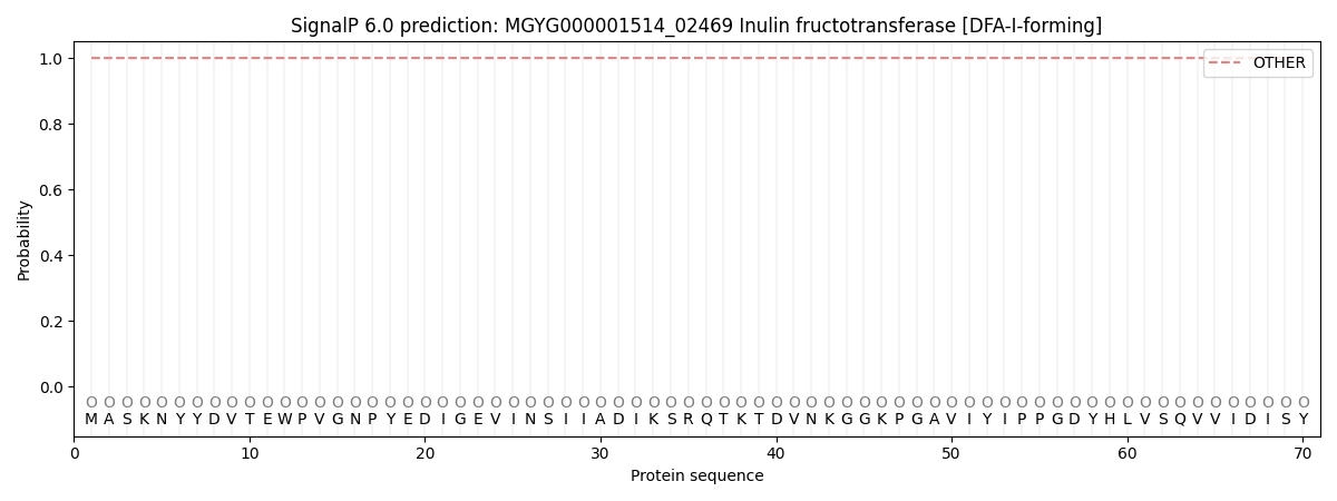
This protein is predicted as OTHER

| Other | SP_Sec_SPI | LIPO_Sec_SPII | TAT_Tat_SPI | TATLIP_Sec_SPII | PILIN_Sec_SPIII |
|---|---|---|---|---|---|
| 1.000071 | 0.000001 | 0.000000 | 0.000000 | 0.000000 | 0.000000 |
