You are browsing environment: HUMAN GUT
CAZyme Information: MGYG000000389_00877
You are here: Home > Sequence: MGYG000000389_00877
Basic Information |
Genomic context |
Full Sequence |
Enzyme annotations |
CAZy signature domains |
CDD domains |
CAZyme hits |
PDB hits |
Swiss-Prot hits |
SignalP and Lipop annotations |
TMHMM annotations
Basic Information help
| Species | Roseburia sp900550935 | |||||||||||
|---|---|---|---|---|---|---|---|---|---|---|---|---|
| Lineage | Bacteria; Firmicutes_A; Clostridia; Lachnospirales; Lachnospiraceae; Roseburia; Roseburia sp900550935 | |||||||||||
| CAZyme ID | MGYG000000389_00877 | |||||||||||
| CAZy Family | GT4 | |||||||||||
| CAZyme Description | Glycosyltransferase Gtf1 | |||||||||||
| CAZyme Property |
|
|||||||||||
| Genome Property |
|
|||||||||||
| Gene Location | Start: 142676; End: 144736 Strand: + | |||||||||||
CDD Domains download full data without filtering help
| Cdd ID | Domain | E-Value | qStart | qEnd | sStart | sEnd | Domain Description |
|---|---|---|---|---|---|---|---|
| cd03808 | GT4_CapM-like | 1.37e-76 | 302 | 675 | 1 | 357 | capsular polysaccharide biosynthesis glycosyltransferase CapM and similar proteins. This family is most closely related to the GT4 family of glycosyltransferases. CapM in Staphylococcus aureus is required for the synthesis of type 1 capsular polysaccharides. |
| cd05232 | UDP_G4E_4_SDR_e | 5.79e-60 | 3 | 295 | 1 | 300 | UDP-glucose 4 epimerase, subgroup 4, extended (e) SDRs. UDP-glucose 4 epimerase (aka UDP-galactose-4-epimerase), is a homodimeric extended SDR. It catalyzes the NAD-dependent conversion of UDP-galactose to UDP-glucose, the final step in Leloir galactose synthesis. This subgroup is comprised of bacterial proteins, and includes the Staphylococcus aureus capsular polysaccharide Cap5N, which may have a role in the synthesis of UDP-N-acetyl-d-fucosamine. This subgroup has the characteristic active site tetrad and NAD-binding motif of the extended SDRs. Extended SDRs are distinct from classical SDRs. In addition to the Rossmann fold (alpha/beta folding pattern with a central beta-sheet) core region typical of all SDRs, extended SDRs have a less conserved C-terminal extension of approximately 100 amino acids. Extended SDRs are a diverse collection of proteins, and include isomerases, epimerases, oxidoreductases, and lyases; they typically have a TGXXGXXG cofactor binding motif. SDRs are a functionally diverse family of oxidoreductases that have a single domain with a structurally conserved Rossmann fold, an NAD(P)(H)-binding region, and a structurally diverse C-terminal region. Sequence identity between different SDR enzymes is typically in the 15-30% range; they catalyze a wide range of activities including the metabolism of steroids, cofactors, carbohydrates, lipids, aromatic compounds, and amino acids, and act in redox sensing. Classical SDRs have an TGXXX[AG]XG cofactor binding motif and a YXXXK active site motif, with the Tyr residue of the active site motif serving as a critical catalytic residue (Tyr-151, human 15-hydroxyprostaglandin dehydrogenase numbering). In addition to the Tyr and Lys, there is often an upstream Ser and/or an Asn, contributing to the active site; while substrate binding is in the C-terminal region, which determines specificity. The standard reaction mechanism is a 4-pro-S hydride transfer and proton relay involving the conserved Tyr and Lys, a water molecule stabilized by Asn, and nicotinamide. Atypical SDRs generally lack the catalytic residues characteristic of the SDRs, and their glycine-rich NAD(P)-binding motif is often different from the forms normally seen in classical or extended SDRs. Complex (multidomain) SDRs such as ketoreductase domains of fatty acid synthase have a GGXGXXG NAD(P)-binding motif and an altered active site motif (YXXXN). Fungal type ketoacyl reductases have a TGXXXGX(1-2)G NAD(P)-binding motif. |
| cd03801 | GT4_PimA-like | 1.36e-46 | 321 | 680 | 24 | 366 | phosphatidyl-myo-inositol mannosyltransferase. This family is most closely related to the GT4 family of glycosyltransferases and named after PimA in Propionibacterium freudenreichii, which is involved in the biosynthesis of phosphatidyl-myo-inositol mannosides (PIM) which are early precursors in the biosynthesis of lipomannans (LM) and lipoarabinomannans (LAM), and catalyzes the addition of a mannosyl residue from GDP-D-mannose (GDP-Man) to the position 2 of the carrier lipid phosphatidyl-myo-inositol (PI) to generate a phosphatidyl-myo-inositol bearing an alpha-1,2-linked mannose residue (PIM1). Glycosyltransferases catalyze the transfer of sugar moieties from activated donor molecules to specific acceptor molecules, forming glycosidic bonds. The acceptor molecule can be a lipid, a protein, a heterocyclic compound, or another carbohydrate residue. This group of glycosyltransferases is most closely related to the previously defined glycosyltransferase family 1 (GT1). The members of this family may transfer UDP, ADP, GDP, or CMP linked sugars. The diverse enzymatic activities among members of this family reflect a wide range of biological functions. The protein structure available for this family has the GTB topology, one of the two protein topologies observed for nucleotide-sugar-dependent glycosyltransferases. GTB proteins have distinct N- and C- terminal domains each containing a typical Rossmann fold. The two domains have high structural homology despite minimal sequence homology. The large cleft that separates the two domains includes the catalytic center and permits a high degree of flexibility. The members of this family are found mainly in certain bacteria and archaea. |
| COG0438 | RfaB | 4.56e-35 | 315 | 682 | 21 | 377 | Glycosyltransferase involved in cell wall bisynthesis [Cell wall/membrane/envelope biogenesis]. |
| cd03819 | GT4_WavL-like | 2.76e-33 | 353 | 631 | 42 | 308 | Vibrio cholerae WavL and similar sequences. This family is most closely related to the GT4 family of glycosyltransferases. WavL in Vibrio cholerae has been shown to be involved in the biosynthesis of the lipopolysaccharide core. |
CAZyme Hits help
| Hit ID | E-Value | Query Start | Query End | Hit Start | Hit End |
|---|---|---|---|---|---|
| VCV20929.1 | 0.0 | 1 | 683 | 1 | 688 |
| CBL07786.1 | 1.75e-172 | 302 | 681 | 4 | 383 |
| QUO31262.1 | 1.82e-154 | 3 | 681 | 317 | 1011 |
| APC41948.1 | 1.53e-110 | 301 | 646 | 2 | 349 |
| QCX14532.1 | 2.94e-110 | 301 | 679 | 2 | 379 |
PDB Hits download full data without filtering help
| Hit ID | E-Value | Query Start | Query End | Hit Start | Hit End | Description |
|---|---|---|---|---|---|---|
| 4XSO_A | 8.00e-15 | 385 | 685 | 89 | 387 | ChainA, Alr3699 protein [Nostoc sp. PCC 7120 = FACHB-418],4XSO_B Chain B, Alr3699 protein [Nostoc sp. PCC 7120 = FACHB-418],4XSP_A Chain A, Alr3699 protein [Nostoc sp. PCC 7120 = FACHB-418],4XSP_B Chain B, Alr3699 protein [Nostoc sp. PCC 7120 = FACHB-418],4XSR_A Chain A, Alr3699 protein [Nostoc sp. PCC 7120 = FACHB-418],4XSR_B Chain B, Alr3699 protein [Nostoc sp. PCC 7120 = FACHB-418],4XSU_A Chain A, Alr3699 protein [Nostoc sp. PCC 7120 = FACHB-418],4XSU_B Chain B, Alr3699 protein [Nostoc sp. PCC 7120 = FACHB-418] |
| 7EC2_A | 1.77e-13 | 409 | 647 | 233 | 462 | ChainA, Glycosyl transferase, group 1 family protein [Staphylococcus aureus subsp. aureus USA300],7EC2_B Chain B, Glycosyl transferase, group 1 family protein [Staphylococcus aureus subsp. aureus USA300] |
| 2JJM_A | 7.57e-10 | 394 | 660 | 101 | 362 | CrystalStructure of a family GT4 glycosyltransferase from Bacillus anthracis ORF BA1558. [Bacillus anthracis str. Ames],2JJM_B Crystal Structure of a family GT4 glycosyltransferase from Bacillus anthracis ORF BA1558. [Bacillus anthracis str. Ames],2JJM_C Crystal Structure of a family GT4 glycosyltransferase from Bacillus anthracis ORF BA1558. [Bacillus anthracis str. Ames],2JJM_D Crystal Structure of a family GT4 glycosyltransferase from Bacillus anthracis ORF BA1558. [Bacillus anthracis str. Ames],2JJM_E Crystal Structure of a family GT4 glycosyltransferase from Bacillus anthracis ORF BA1558. [Bacillus anthracis str. Ames],2JJM_F Crystal Structure of a family GT4 glycosyltransferase from Bacillus anthracis ORF BA1558. [Bacillus anthracis str. Ames],2JJM_G Crystal Structure of a family GT4 glycosyltransferase from Bacillus anthracis ORF BA1558. [Bacillus anthracis str. Ames],2JJM_H Crystal Structure of a family GT4 glycosyltransferase from Bacillus anthracis ORF BA1558. [Bacillus anthracis str. Ames],2JJM_I Crystal Structure of a family GT4 glycosyltransferase from Bacillus anthracis ORF BA1558. [Bacillus anthracis str. Ames],2JJM_J Crystal Structure of a family GT4 glycosyltransferase from Bacillus anthracis ORF BA1558. [Bacillus anthracis str. Ames],2JJM_K Crystal Structure of a family GT4 glycosyltransferase from Bacillus anthracis ORF BA1558. [Bacillus anthracis str. Ames],2JJM_L Crystal Structure of a family GT4 glycosyltransferase from Bacillus anthracis ORF BA1558. [Bacillus anthracis str. Ames] |
| 3MBO_A | 8.17e-10 | 394 | 660 | 121 | 382 | CrystalStructure of the Glycosyltransferase BaBshA bound with UDP and L-malate [Bacillus anthracis],3MBO_B Crystal Structure of the Glycosyltransferase BaBshA bound with UDP and L-malate [Bacillus anthracis],3MBO_C Crystal Structure of the Glycosyltransferase BaBshA bound with UDP and L-malate [Bacillus anthracis],3MBO_D Crystal Structure of the Glycosyltransferase BaBshA bound with UDP and L-malate [Bacillus anthracis],3MBO_E Crystal Structure of the Glycosyltransferase BaBshA bound with UDP and L-malate [Bacillus anthracis],3MBO_F Crystal Structure of the Glycosyltransferase BaBshA bound with UDP and L-malate [Bacillus anthracis],3MBO_G Crystal Structure of the Glycosyltransferase BaBshA bound with UDP and L-malate [Bacillus anthracis],3MBO_H Crystal Structure of the Glycosyltransferase BaBshA bound with UDP and L-malate [Bacillus anthracis] |
| 5D00_A | 1.66e-09 | 377 | 640 | 74 | 349 | Crystalstructure of BshA from B. subtilis complexed with N-acetylglucosaminyl-malate and UMP [Bacillus subtilis subsp. subtilis str. 168],5D00_B Crystal structure of BshA from B. subtilis complexed with N-acetylglucosaminyl-malate and UMP [Bacillus subtilis subsp. subtilis str. 168],5D01_A Crystal structure of BshA from B. subtilis complexed with N-acetylglucosaminyl-malate [Bacillus subtilis subsp. subtilis str. 168],5D01_B Crystal structure of BshA from B. subtilis complexed with N-acetylglucosaminyl-malate [Bacillus subtilis subsp. subtilis str. 168] |
Swiss-Prot Hits download full data without filtering help
| Hit ID | E-Value | Query Start | Query End | Hit Start | Hit End | Description |
|---|---|---|---|---|---|---|
| P71053 | 1.87e-61 | 301 | 679 | 3 | 368 | Putative glycosyltransferase EpsD OS=Bacillus subtilis (strain 168) OX=224308 GN=epsD PE=2 SV=1 |
| Q56623 | 8.70e-18 | 52 | 303 | 74 | 319 | UDP-glucose 4-epimerase OS=Vibrio cholerae OX=666 GN=galE PE=3 SV=1 |
| Q58459 | 2.08e-13 | 352 | 606 | 41 | 304 | Uncharacterized glycosyltransferase MJ1059 OS=Methanocaldococcus jannaschii (strain ATCC 43067 / DSM 2661 / JAL-1 / JCM 10045 / NBRC 100440) OX=243232 GN=MJ1059 PE=3 SV=1 |
| P71055 | 4.18e-11 | 480 | 615 | 178 | 316 | Putative glycosyltransferase EpsF OS=Bacillus subtilis (strain 168) OX=224308 GN=epsF PE=2 SV=1 |
| Q46638 | 1.10e-10 | 509 | 641 | 226 | 368 | Amylovoran biosynthesis glycosyltransferase AmsK OS=Erwinia amylovora OX=552 GN=amsK PE=3 SV=2 |
SignalP and Lipop Annotations help
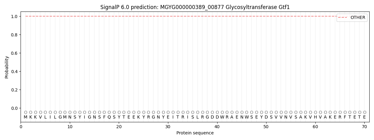
This protein is predicted as OTHER
| Other | SP_Sec_SPI | LIPO_Sec_SPII | TAT_Tat_SPI | TATLIP_Sec_SPII | PILIN_Sec_SPIII |
|---|---|---|---|---|---|
| 1.000080 | 0.000001 | 0.000000 | 0.000000 | 0.000000 | 0.000000 |