You are browsing environment: HUMAN GUT
CAZyme Information: MGYG000000137_01391
You are here: Home > Sequence: MGYG000000137_01391
Basic Information |
Genomic context |
Full Sequence |
Enzyme annotations |
CAZy signature domains |
CDD domains |
CAZyme hits |
PDB hits |
Swiss-Prot hits |
SignalP and Lipop annotations |
TMHMM annotations
Basic Information help
| Species | UBA7160 sp902363665 | |||||||||||
|---|---|---|---|---|---|---|---|---|---|---|---|---|
| Lineage | Bacteria; Firmicutes_A; Clostridia; Lachnospirales; Lachnospiraceae; UBA7160; UBA7160 sp902363665 | |||||||||||
| CAZyme ID | MGYG000000137_01391 | |||||||||||
| CAZy Family | GH13 | |||||||||||
| CAZyme Description | Trehalose synthase/amylase TreS | |||||||||||
| CAZyme Property |
|
|||||||||||
| Genome Property |
|
|||||||||||
| Gene Location | Start: 71263; End: 72810 Strand: - | |||||||||||
CAZyme Signature Domains help
| Family | Start | End | Evalue | family coverage |
|---|---|---|---|---|
| GH13 | 27 | 358 | 1.1e-66 | 0.9264214046822743 |
CDD Domains download full data without filtering help
| Cdd ID | Domain | E-Value | qStart | qEnd | sStart | sEnd | Domain Description |
|---|---|---|---|---|---|---|---|
| cd11348 | AmyAc_2 | 0.0 | 9 | 430 | 1 | 429 | Alpha amylase catalytic domain found in an uncharacterized protein family. The Alpha-amylase family comprises the largest family of glycoside hydrolases (GH), with the majority of enzymes acting on starch, glycogen, and related oligo- and polysaccharides. These proteins catalyze the transformation of alpha-1,4 and alpha-1,6 glucosidic linkages with retention of the anomeric center. The protein is described as having 3 domains: A, B, C. A is a (beta/alpha) 8-barrel; B is a loop between the beta 3 strand and alpha 3 helix of A; C is the C-terminal extension characterized by a Greek key. The majority of the enzymes have an active site cleft found between domains A and B where a triad of catalytic residues (Asp, Glu and Asp) performs catalysis. Other members of this family have lost the catalytic activity as in the case of the human 4F2hc, or only have 2 residues that serve as the catalytic nucleophile and the acid/base, such as Thermus A4 beta-galactosidase with 2 Glu residues (GH42) and human alpha-galactosidase with 2 Asp residues (GH31). The catalytic triad (DED) is not present here. The family members are quite extensive and include: alpha amylase, maltosyltransferase, cyclodextrin glycotransferase, maltogenic amylase, neopullulanase, isoamylase, 1,4-alpha-D-glucan maltotetrahydrolase, 4-alpha-glucotransferase, oligo-1,6-glucosidase, amylosucrase, sucrose phosphorylase, and amylomaltase. |
| cd11333 | AmyAc_SI_OligoGlu_DGase | 3.73e-113 | 8 | 433 | 3 | 428 | Alpha amylase catalytic domain found in Sucrose isomerases, oligo-1,6-glucosidase (also called isomaltase; sucrase-isomaltase; alpha-limit dextrinase), dextran glucosidase (also called glucan 1,6-alpha-glucosidase), and related proteins. The sucrose isomerases (SIs) Isomaltulose synthase (EC 5.4.99.11) and Trehalose synthase (EC 5.4.99.16) catalyze the isomerization of sucrose and maltose to produce isomaltulose and trehalulose, respectively. Oligo-1,6-glucosidase (EC 3.2.1.10) hydrolyzes the alpha-1,6-glucosidic linkage of isomaltooligosaccharides, pannose, and dextran. Unlike alpha-1,4-glucosidases (EC 3.2.1.20), it fails to hydrolyze the alpha-1,4-glucosidic bonds of maltosaccharides. Dextran glucosidase (DGase, EC 3.2.1.70) hydrolyzes alpha-1,6-glucosidic linkages at the non-reducing end of panose, isomaltooligosaccharides and dextran to produce alpha-glucose.The common reaction chemistry of the alpha-amylase family enzymes is based on a two-step acid catalytic mechanism that requires two critical carboxylates: one acting as a general acid/base (Glu) and the other as a nucleophile (Asp). Both hydrolysis and transglycosylation proceed via the nucleophilic substitution reaction between the anomeric carbon, C1 and a nucleophile. Both enzymes contain the three catalytic residues (Asp, Glu and Asp) common to the alpha-amylase family as well as two histidine residues which are predicted to be critical to binding the glucose residue adjacent to the scissile bond in the substrates. The Alpha-amylase family comprises the largest family of glycoside hydrolases (GH), with the majority of enzymes acting on starch, glycogen, and related oligo- and polysaccharides. These proteins catalyze the transformation of alpha-1,4 and alpha-1,6 glucosidic linkages with retention of the anomeric center. The protein is described as having 3 domains: A, B, C. A is a (beta/alpha) 8-barrel; B is a loop between the beta 3 strand and alpha 3 helix of A; C is the C-terminal extension characterized by a Greek key. The majority of the enzymes have an active site cleft found between domains A and B where a triad of catalytic residues performs catalysis. Other members of this family have lost the catalytic activity as in the case of the human 4F2hc, or only have 2 residues that serve as the catalytic nucleophile and the acid/base, such as Thermus A4 beta-galactosidase with 2 Glu residues (GH42) and human alpha-galactosidase with 2 Asp residues (GH31). The family members are quite extensive and include: alpha amylase, maltosyltransferase, cyclodextrin glycotransferase, maltogenic amylase, neopullulanase, isoamylase, 1,4-alpha-D-glucan maltotetrahydrolase, 4-alpha-glucotransferase, oligo-1,6-glucosidase, amylosucrase, sucrose phosphorylase, and amylomaltase. |
| cd11316 | AmyAc_bac2_AmyA | 1.47e-109 | 8 | 440 | 1 | 403 | Alpha amylase catalytic domain found in bacterial Alpha-amylases (also called 1,4-alpha-D-glucan-4-glucanohydrolase). AmyA (EC 3.2.1.1) catalyzes the hydrolysis of alpha-(1,4) glycosidic linkages of glycogen, starch, related polysaccharides, and some oligosaccharides. This group includes Chloroflexi, Dictyoglomi, and Fusobacteria. The Alpha-amylase family comprises the largest family of glycoside hydrolases (GH), with the majority of enzymes acting on starch, glycogen, and related oligo- and polysaccharides. These proteins catalyze the transformation of alpha-1,4 and alpha-1,6 glucosidic linkages with retention of the anomeric center. The protein is described as having 3 domains: A, B, C. A is a (beta/alpha) 8-barrel; B is a loop between the beta 3 strand and alpha 3 helix of A; C is the C-terminal extension characterized by a Greek key. The majority of the enzymes have an active site cleft found between domains A and B where a triad of catalytic residues (Asp, Glu and Asp) performs catalysis. Other members of this family have lost the catalytic activity as in the case of the human 4F2hc, or only have 2 residues that serve as the catalytic nucleophile and the acid/base, such as Thermus A4 beta-galactosidase with 2 Glu residues (GH42) and human alpha-galactosidase with 2 Asp residues (GH31). The family members are quite extensive and include: alpha amylase, maltosyltransferase, cyclodextrin glycotransferase, maltogenic amylase, neopullulanase, isoamylase, 1,4-alpha-D-glucan maltotetrahydrolase, 4-alpha-glucotransferase, oligo-1,6-glucosidase, amylosucrase, sucrose phosphorylase, and amylomaltase. |
| cd11331 | AmyAc_OligoGlu_like | 1.99e-100 | 3 | 441 | 1 | 450 | Alpha amylase catalytic domain found in oligo-1,6-glucosidase (also called isomaltase; sucrase-isomaltase; alpha-limit dextrinase) and related proteins. Oligo-1,6-glucosidase (EC 3.2.1.10) hydrolyzes the alpha-1,6-glucosidic linkage of isomalto-oligosaccharides, pannose, and dextran. Unlike alpha-1,4-glucosidases (EC 3.2.1.20), it fails to hydrolyze the alpha-1,4-glucosidic bonds of maltosaccharides. The Alpha-amylase family comprises the largest family of glycoside hydrolases (GH), with the majority of enzymes acting on starch, glycogen, and related oligo- and polysaccharides. These proteins catalyze the transformation of alpha-1,4 and alpha-1,6 glucosidic linkages with retention of the anomeric center. The protein is described as having 3 domains: A, B, C. A is a (beta/alpha) 8-barrel; B is a loop between the beta 3 strand and alpha 3 helix of A; C is the C-terminal extension characterized by a Greek key. The majority of the enzymes have an active site cleft found between domains A and B where a triad of catalytic residues (Asp, Glu and Asp) performs catalysis. Other members of this family have lost the catalytic activity as in the case of the human 4F2hc, or only have 2 residues that serve as the catalytic nucleophile and the acid/base, such as Thermus A4 beta-galactosidase with 2 Glu residues (GH42) and human alpha-galactosidase with 2 Asp residues (GH31). The family members are quite extensive and include: alpha amylase, maltosyltransferase, cyclodextrin glycotransferase, maltogenic amylase, neopullulanase, isoamylase, 1,4-alpha-D-glucan maltotetrahydrolase, 4-alpha-glucotransferase, oligo-1,6-glucosidase, amylosucrase, sucrose phosphorylase, and amylomaltase. |
| cd11334 | AmyAc_TreS | 1.33e-99 | 4 | 431 | 1 | 447 | Alpha amylase catalytic domain found in Trehalose synthetase. Trehalose synthetase (TreS) catalyzes the reversible interconversion of trehalose and maltose. The enzyme catalyzes the reaction in both directions, but the preferred substrate is maltose. Glucose is formed as a by-product of this reaction. It is believed that the catalytic mechanism may involve the cutting of the incoming disaccharide and transfer of a glucose to an enzyme-bound glucose. This enzyme also catalyzes production of a glucosamine disaccharide from maltose and glucosamine. The Alpha-amylase family comprises the largest family of glycoside hydrolases (GH), with the majority of enzymes acting on starch, glycogen, and related oligo- and polysaccharides. These proteins catalyze the transformation of alpha-1,4 and alpha-1,6 glucosidic linkages with retention of the anomeric center. The protein is described as having 3 domains: A, B, C. A is a (beta/alpha) 8-barrel; B is a loop between the beta 3 strand and alpha 3 helix of A; C is the C-terminal extension characterized by a Greek key. The majority of the enzymes have an active site cleft found between domains A and B where a triad of catalytic residues (Asp, Glu and Asp) performs catalysis. Other members of this family have lost the catalytic activity as in the case of the human 4F2hc, or only have 2 residues that serve as the catalytic nucleophile and the acid/base, such as Thermus A4 beta-galactosidase with 2 Glu residues (GH42) and human alpha-galactosidase with 2 Asp residues (GH31). The family members are quite extensive and include: alpha amylase, maltosyltransferase, cyclodextrin glycotransferase, maltogenic amylase, neopullulanase, isoamylase, 1,4-alpha-D-glucan maltotetrahydrolase, 4-alpha-glucotransferase, oligo-1,6-glucosidase, amylosucrase, sucrose phosphorylase, and amylomaltase. |
CAZyme Hits help
| Hit ID | E-Value | Query Start | Query End | Hit Start | Hit End |
|---|---|---|---|---|---|
| QOL32207.1 | 1.76e-249 | 1 | 512 | 1 | 514 |
| QTL77951.1 | 1.46e-233 | 1 | 514 | 1 | 531 |
| QTL79841.1 | 1.46e-233 | 1 | 514 | 1 | 531 |
| BAQ26095.1 | 1.19e-232 | 1 | 514 | 1 | 531 |
| ADB08796.1 | 1.19e-232 | 1 | 514 | 1 | 531 |
PDB Hits download full data without filtering help
| Hit ID | E-Value | Query Start | Query End | Hit Start | Hit End | Description |
|---|---|---|---|---|---|---|
| 4TVU_A | 6.25e-73 | 3 | 485 | 9 | 515 | Crystalstructure of trehalose synthase from Deinococcus radiodurans reveals a closed conformation for catalysis of the intramolecular isomerization [Deinococcus radiodurans R1],4TVU_B Crystal structure of trehalose synthase from Deinococcus radiodurans reveals a closed conformation for catalysis of the intramolecular isomerization [Deinococcus radiodurans R1],4TVU_C Crystal structure of trehalose synthase from Deinococcus radiodurans reveals a closed conformation for catalysis of the intramolecular isomerization [Deinococcus radiodurans R1],4TVU_D Crystal structure of trehalose synthase from Deinococcus radiodurans reveals a closed conformation for catalysis of the intramolecular isomerization [Deinococcus radiodurans R1],4TVU_E Crystal structure of trehalose synthase from Deinococcus radiodurans reveals a closed conformation for catalysis of the intramolecular isomerization [Deinococcus radiodurans R1],4TVU_F Crystal structure of trehalose synthase from Deinococcus radiodurans reveals a closed conformation for catalysis of the intramolecular isomerization [Deinococcus radiodurans R1],4TVU_G Crystal structure of trehalose synthase from Deinococcus radiodurans reveals a closed conformation for catalysis of the intramolecular isomerization [Deinococcus radiodurans R1],4TVU_H Crystal structure of trehalose synthase from Deinococcus radiodurans reveals a closed conformation for catalysis of the intramolecular isomerization [Deinococcus radiodurans R1] |
| 4WF7_A | 6.25e-73 | 3 | 485 | 9 | 515 | Crystalstructures of trehalose synthase from Deinococcus radiodurans reveal that a closed conformation is involved in the intramolecular isomerization catalysis [Deinococcus radiodurans R1],4WF7_B Crystal structures of trehalose synthase from Deinococcus radiodurans reveal that a closed conformation is involved in the intramolecular isomerization catalysis [Deinococcus radiodurans R1],4WF7_C Crystal structures of trehalose synthase from Deinococcus radiodurans reveal that a closed conformation is involved in the intramolecular isomerization catalysis [Deinococcus radiodurans R1],4WF7_D Crystal structures of trehalose synthase from Deinococcus radiodurans reveal that a closed conformation is involved in the intramolecular isomerization catalysis [Deinococcus radiodurans R1] |
| 5GTW_A | 8.75e-73 | 3 | 485 | 9 | 515 | TheN253R mutant structures of trehalose synthase from Deinococcus radiodurans display two different active-site conformations [Deinococcus radiodurans R1],5GTW_B The N253R mutant structures of trehalose synthase from Deinococcus radiodurans display two different active-site conformations [Deinococcus radiodurans R1],5GTW_C The N253R mutant structures of trehalose synthase from Deinococcus radiodurans display two different active-site conformations [Deinococcus radiodurans R1],5GTW_D The N253R mutant structures of trehalose synthase from Deinococcus radiodurans display two different active-site conformations [Deinococcus radiodurans R1] |
| 5YKB_A | 8.75e-73 | 3 | 485 | 9 | 515 | TheN253F mutant structure of trehalose synthase from Deinococcus radiodurans reveals an open active-site conformation [Deinococcus radiodurans R1],5YKB_B The N253F mutant structure of trehalose synthase from Deinococcus radiodurans reveals an open active-site conformation [Deinococcus radiodurans R1],5YKB_C The N253F mutant structure of trehalose synthase from Deinococcus radiodurans reveals an open active-site conformation [Deinococcus radiodurans R1],5YKB_D The N253F mutant structure of trehalose synthase from Deinococcus radiodurans reveals an open active-site conformation [Deinococcus radiodurans R1] |
| 5H2T_A | 3.31e-72 | 4 | 458 | 22 | 498 | Structureof trehalose synthase [Thermomonospora curvata DSM 43183],5H2T_B Structure of trehalose synthase [Thermomonospora curvata DSM 43183],5H2T_C Structure of trehalose synthase [Thermomonospora curvata DSM 43183],5H2T_D Structure of trehalose synthase [Thermomonospora curvata DSM 43183],5H2T_E Structure of trehalose synthase [Thermomonospora curvata DSM 43183],5H2T_F Structure of trehalose synthase [Thermomonospora curvata DSM 43183],5H2T_G Structure of trehalose synthase [Thermomonospora curvata DSM 43183],5H2T_H Structure of trehalose synthase [Thermomonospora curvata DSM 43183] |
Swiss-Prot Hits download full data without filtering help
| Hit ID | E-Value | Query Start | Query End | Hit Start | Hit End | Description |
|---|---|---|---|---|---|---|
| A0R6E0 | 4.81e-67 | 2 | 458 | 33 | 511 | Trehalose synthase/amylase TreS OS=Mycolicibacterium smegmatis (strain ATCC 700084 / mc(2)155) OX=246196 GN=treS PE=1 SV=1 |
| P29094 | 1.92e-65 | 4 | 490 | 5 | 526 | Oligo-1,6-glucosidase OS=Parageobacillus thermoglucosidasius OX=1426 GN=malL PE=1 SV=1 |
| P9WQ19 | 6.09e-64 | 4 | 456 | 43 | 517 | Trehalose synthase/amylase TreS OS=Mycobacterium tuberculosis (strain ATCC 25618 / H37Rv) OX=83332 GN=treS PE=1 SV=1 |
| P9WQ18 | 6.09e-64 | 4 | 456 | 43 | 517 | Trehalose synthase/amylase TreS OS=Mycobacterium tuberculosis (strain CDC 1551 / Oshkosh) OX=83331 GN=treS PE=3 SV=1 |
| O06994 | 5.41e-63 | 1 | 477 | 1 | 518 | Oligo-1,6-glucosidase 1 OS=Bacillus subtilis (strain 168) OX=224308 GN=malL PE=1 SV=1 |
SignalP and Lipop Annotations help
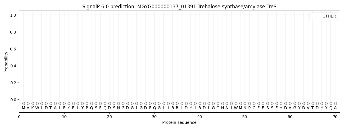
This protein is predicted as OTHER

| Other | SP_Sec_SPI | LIPO_Sec_SPII | TAT_Tat_SPI | TATLIP_Sec_SPII | PILIN_Sec_SPIII |
|---|---|---|---|---|---|
| 1.000055 | 0.000000 | 0.000000 | 0.000000 | 0.000000 | 0.000000 |
